
Clinical History
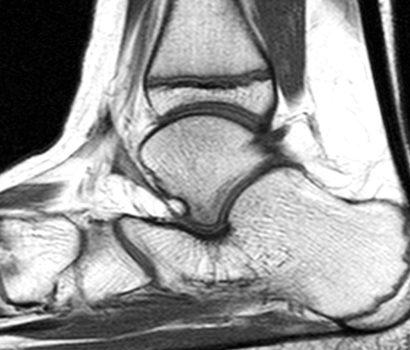
A 9-year-old boy with a rigid flatfoot presents with hindfoot pain. (1A, 1B) Sagittal T1-weighted, (1C) sagittal fat-suppressed, fluid-sensitive and (1D) coronal fat-suppressed, fluid-sensitive images are provided. What are the findings? What is your diagnosis?



Findings

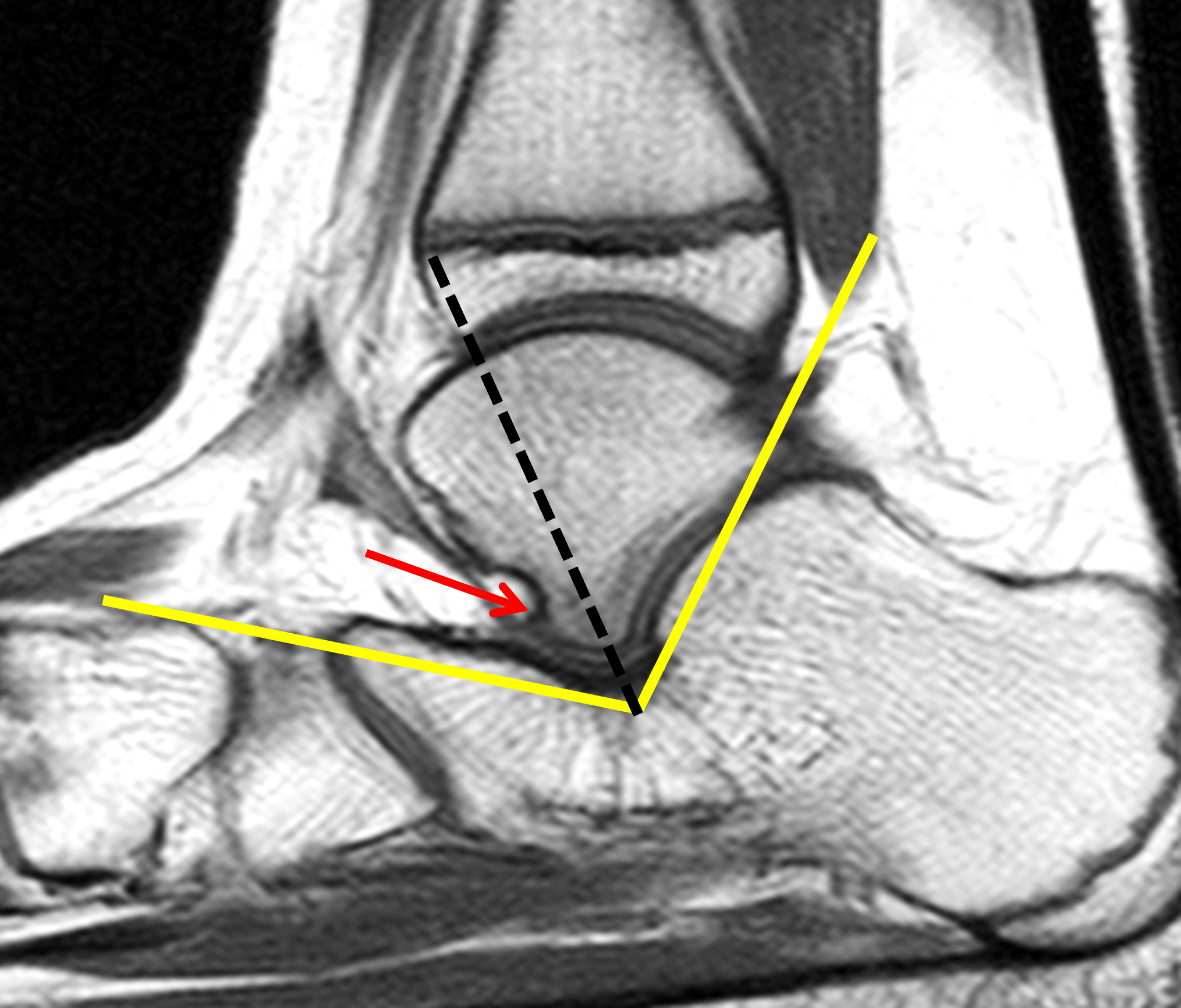


Figure 2: (2A) Sagittal T1-weighted image through the medial hindfoot shows plantar flexion of the talus with its long axis (dotted black line) pointing downward rather than being aligned with the first metatarsal. While foot malalignment may be underestimated on (nonweightbearing) MRI, this talar orientation suggests pes planus. Note the small dorsal talar beak (red arrow). No evidence of middle facet coalition. (2B) Sagittal T1-weighted image through the lateral hindfoot shows an accessory anterolateral talar facet (AALTF, red arrow), which abuts the calcaneus at the angle of Gissane. The lateral process of the talus should not extend anterior to a bisector of the Gissane angle (dotted line). Mild cortical thickening of the calcaneus adjacent to the AALTF may be due to increased mechanical stress. No evidence of posterior facet coalition. (2C) Sagittal fat-suppressed, fluid-sensitive image shows shows subjacent bone marrow edema-like changes (asterisks) around the AALTF. (2D) Coronal fat-suppressed, fluid-sensitive image shows lateral shift of the calcaneus (C) with respect to the talus (T), consistent with hindfoot valgus. Note the diffuse marrow edema (asterisks) in the lateral hindfoot at the AALTF.
Diagnosis
Rigid flatfoot with an accessory anterolateral talar facet (AALTF) and hindfoot impingement.
Introduction
Pediatric flatfoot should not be considered a variant of acquired adult flat foot deformity. The common, flexible-type flatfoot in children is typically asymptomatic and benign without risk for progressive deformity or future disability. However, several variants or subtypes of pediatric flatfoot should be recognized and treated to prevent permanent impairment. Recognize that the diagnosis of flatfoot (pes planus) is controversial because there is no general agreement on the clinical or radiographic criteria. Traditionally, clinicians have diagnosed flatfoot by observing a low or absent arch during weight-bearing, but the mobility of the joints and tendons are more important than the shape of the foot for patient management.1,2
Normative radiographic measurements and angular relationships are available for adults and children, but normal ranges vary widely by age.3 Accurate measurement is limited by factors such as patient positioning, degree of weightbearing, radiographic exposure, and intra- and interobserver variability. These limitations call into question the reliability and clinical relevance of radiographic measurements.4 Radiographs alone should not direct treatment. An evaluation that includes symptoms, flexibility and function is essential.5
Comprehensive evaluation of flatfoot is only needed if there are symptoms such as gait issues, regional pain and stiffness. Imaging work-up typically starts with weight-bearing radiographs. If there is a strong suspicion of an underlying structural abnormality such as tarsal coalition or an accessory anterolateral talar facet (AALTF), further diagnostic work-up with CT or MRI is warranted.6
Anatomy and Pathophysiology
The longitudinal arch of the foot has a medial and lateral component. The medial longitudinal arch consists of the calcaneus, talus, tarsal navicular bone, the three cuneiform bones, and the first three metatarsals. The spring ligament, plantar aponeurosis, and muscles and tendons – primarily the posterior tibial and flexor hallucis longus – are crucial supporting structures. The lateral longitudinal arch is formed by the calcaneus, cuboid, and the fourth and fifth metatarsal bones. Key supporting structures include the long and short plantar ligaments and the peroneus longus and brevis tendons.7
The transverse arch of the foot consists of the three cuneiforms, the cuboid, and the bases of the five metatarsals. The transverse arch has two parts, proximal (at the tarsal level) and distal (at the metatarsal head level), and is supported by the deep transverse metatarsal ligaments, plantar ligaments, dorsal/interosseous ligaments, peroneus longus and posterior tibialis tendons, and intrinsic foot muscles.
The flat appearance of the plantar arch in infants is normal and is caused mainly by the prominent plantar fat pad. As the child grows and starts walking, the feet remain relatively flat due to remaining normal plantar fat and physiologic laxity of the ligaments.8,9 At approximately 5 years of age, the arch has risen spontaneously in most children, and between the ages of 7 and 10 years, arch development is complete. In some children the feet remain relatively flat, but only a small number will have related symptoms. The true prevalence of flatfoot remains unknown because standardized diagnostic criteria are lacking.
Genetic factors play a major role in pediatric flatfoot because the shape of the bones and the degree of ligament laxity are partly inherited. Tightness of the Achilles tendon and calf muscles in the setting of flexible flatfoot is often associated with pain and disability.10 Congenital factors also play a role in calcaneovalgus and vertical talus.
Childhood obesity carries a high risk of abnormal development of the plantar arch during growth due to extra body weight and abnormal mechanical strain on the foot,9 and the deformity will often persist into adulthood. Obese children also have increased thickness of the plantar fat, which could make the foot appear flatter.
Genetic syndromes, such as Ehlers-Danlos syndrome and Marfan syndrome, can be associated with flatfoot deformity at a young age due to abnormal connective tissues. Patients with Down syndrome have a high frequency (58%) of flatfoot deformity due to hypotonia and increased ligamentous laxity.11
Late-presenting rigid flatfoot is generally considered an acquired deformity and is often caused by tarsal coalition. An autosomal dominant inheritance pattern has been observed in cases of tarsal coalition.12
In chronic neuromuscular disorders such as cerebral palsy, foot malignment resembling flatfoot may develop as a result of muscular imbalance and chronic spasticity.13 Rarely, trauma can lead to acquired flatfoot in children, including fractures and spring ligament tears.14
Clinical Evaluation
Flatfoot deformity, if clinically significant, usually comes to attention at the time the child has reached school age (6-12 years) due to gait disturbance and pain. Physical examination includes inspection, the tiptoe test (Jack’s test), analysis of gait and range of motion, and observation of the wear patterns on shoes.15
In a flexible flatfoot, increased laxity of the foot ligaments allows the medial longitudinal arch to flatten when the child stands. The whole foot may press against the floor during weight-bearing. The arch is visible in seated or tiptoe position (non-weightbearing), and the general range of motion is preserved. Contracture of the Achilles tendon and calf muscles is found in association with flexible flatfoot in 25% and is thought to be a contributing factor to pain in flexible flatfoot.
The rigid flatfoot, encountered primarily with congenital vertical talus and sometimes with tarsal coalition, is characterized by restricted motion of the subtalar joint. The underlying structural abnormalities in these entities keep the foot flat in all positions, and the range of motion is limited. A classic presentation of tarsal coalition is peroneal spastic flatfoot, which is a painful condition caused by chronic spasm and/or adaptive shortening of the peroneal muscles, resulting in foot pronation and abduction.16
Imaging Evaluation
Standard ankle radiographic projections include AP, lateral and mortise views with weight bearing; standard foot radiographs include AP (i.e., dorsoplantar) and lateral views with weight bearing.5 The lateral view of the foot should ideally be taken with the foot in dorsiflexion. In instances where patients are unable to bear weight, pressure can be applied on the plantar surface of the foot by using a wooden or plastic board to simulate weightbearing. For assessment of the subtalar range of motion and overall rigidity of the foot, lateral radiographs with the foot in maximum plantarflexion and dorsiflexion may be helpful. Oblique views of the foot are not mandatory in the initial work up or follow up evaluation of foot deformities.
Weight-bearing radiographs in children with flatfoot show varying degrees of hindfoot valgus and plantar flexion (a more vertical position) of the talus,6 often within normal limits. The calcaneus and metatarsals are often more horizontally oriented. Additional findings that are not always present or obvious include foot eversion and lateral shift of the navicular with respect to the talus. A complete analysis of normal and abnormal foot and ankle alignment is complex17 and is beyond the scope of this Web Clinic.
On lateral weight-bearing radiographs, the following angular relationships are evaluated (Figure 3A): The talar-first metatarsal (Meary) angle, the calcaneal inclination angle, and the calcaneal-fifth metatarsal angle. On AP weight-bearing radiographs (Figure 3B), the talocalcaneal (Kite angle), the talus-first metatarsal axis, and the talonavicular coverage angle are assessed. The cyma line is a curved line joining the talonavicular and calcaneocuboid joints and serves as a radiographic indicator of midtarsal alignment on lateral and AP views. It is frequently altered in pediatric flatfoot but may also be abnormal with Chopart injuries and fractures.18
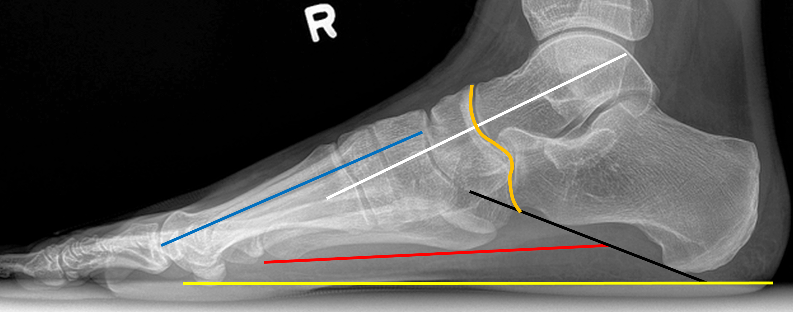

Figure 3: Weightbearing foot radiographs of a young adult with normal alignment. (3A) Lateral view. The midtalar line (white line) bisects the anterior and posterior talus and should parallel or overlap with the long axis of the first metatarsal (blue line). Meary’s angle describes the angle between the talar and first metatarsal long axes. Calcaneal inclination (calcaneal pitch) is the angle between the plantar surface of the calcaneus (black line) and the horizontal supporting surface (yellow line), which is decreased with pes planus. The calcaneal-fifth metatarsal (arch angle) angle is formed between the calcaneal inclination axis (black line) and a line drawn along the inferior edge of the fifth metatarsal (red line). The normal cyma line is a smooth S-shaped curve formed by the talonavicular and calcaneocuboid joints (curved orange line). (3B) AP (dorsoplantar) view. A line through the long axis of the talus (red line) should pass through or just medial to the base of the first metatarsal. A line through the mid calcaneus (black line) should intersect the base of the fourth metatarsal. The talocalcaneal (Kite’s) angle is the angle between these two axes. Talonavicular coverage is assessed by measuring the angular relationship between the talar head and the adjacent navicular. The cyma line (curved orange line) is smooth and continuous.
Additional views of the foot, such as the oblique and the Harris-Beath views, aid in the detection of coalitions.
Following standard initial radiographs, CT and/or MRI are performed to evaluate suspected underlying structural abnormalities. MRI of the foot and ankle may play a role in selected patients under the age of 18 with a history of flatfoot. In a recent study,19 MRI provided additional relevant diagnostic information in 19 of 81 feet (23.5%) including improved detection of talocalcaneal and calcaneonavicular coalitions. The authors also concluded that MRI is the gold-standard for detection of non-osseous (fibrous and cartilaginous) tarsal coalition. Furthermore, in patients with flatfoot and type II accessory navicular, two entities that are frequently linked, MRI may show focal marrow edema, which correlates with symptoms, and is helpful for clinical decision-making.20
Zero echo time (ZTE) MRI improves visualization of cortical bone and produces CT-like images without ionizing radiation. This emerging technique may reduce the need for conventional CT in evaluating pediatric musculoskeletal disorders.21
Imaging Findings: Subtypes and Associated Findings
Physiologic flatfoot
Imaging is not recommended in the setting of asymptomatic flatfoot (flexible and painless), but when performed, it is important to recognize that the longitudinal arch is not fully developed until the age of about 10 years, and the standard radiographic measurements used in older children and adults do not apply (Figure 4). Normal hindfoot-forefoot angular relationships have been established for different age groups up to 9 years, but the intervals for each age group are wide and the radiographic measurements may be inaccurate.

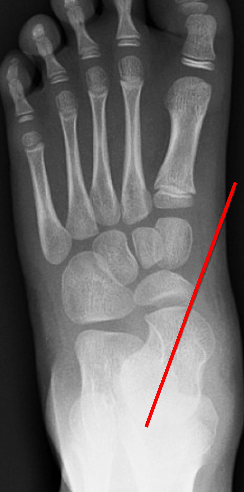
Figure 4: Normal weightbearing foot radiographs in a 5 year old. (4A) Lateral view. The mid talar line (red line) passes inferior to the first metatarsal, which is within normal limits at this age. (4B) AP (dorsoplantar) view. The mid talar line (red line) passes medial to the first metatarsal, within normal limits at this age. The talocalcaneal angle is preserved.
Congenital Calcaneovalgus
30-50% of newborns have varying degrees of a benign flexible foot deformity, known as congenital calcaneovalgus, which is frequently bilateral and likely related to intrauterine crowding. The foot is curved and bent upward and outward, with the forefoot abducted. The talus is in plantar flexion, similar to congenital vertical talus, but the position of the calcaneus differs between the two entities. In congenital calcaneovalgus, the calcaneus is in dorsiflexion (Figure 5), while in congenital vertical talus, the calcaneus is in plantar flexion (equinus) with respect to the tibia.22 Because of its benign course, congenital calcaneovalgus is rarely imaged.

Figure 5: Infant with congenital calcaneovalgus, a benign flexible flatfoot deformity. Lateral, non-weightbearing radiograph shows plantar flexion of the talus (yellow line) with flattening of the arch. The anterior end of the calcaneus is in dorsiflexion with respect to the ankle (red line = long axis of the calcaneus, black line = long axis of the tibia), which differs from congenital vertical talus, where the calcaneus is in plantar flexion (equinus).
Congenital Vertical Talus
The underlying abnormality of a rigid congenital flat foot is a vertical talus (Figure 6), a condition of unclear etiology with an incidence of 1 in 10,000 births.23 “Rocker-bottom deformity” is a clinical term that does not always imply a congenital vertical talus. A genetic predisposition has been proposed. Additionally, an association with syndromes, such as trisomies, Freeman-Sheldon syndrome, myelomeningocele, neurofibromatosis and arthrogryposis) is present in at least 50% of cases. Abnormal position of tendons at the ankle may prevent normal plantar flexion of the foot, and a short Achilles tendon keeps the calcaneus in plantar flexion (equinus). There is also dorsal dislocation of the navicular bone.


Figure 6. Congenital vertical talus and rigid flatfoot deformity in two patients. (6A) Lateral view of the foot in an infant shows vertical orientation of the talus, almost parallel with the tibia. The talocalcaneal angle is increased and the calcaneus is in equinus (compare with Figure 5). The navicular bone is not yet ossified at this age. (6B) Corresponding AP view shows medial orientation of the talus and lateral displacement of the calcaneus with hindfoot valgus. The forefoot is mildly everted with decreased convergence of the metatarsal bases. (6C) Non-weightbearing lateral view of the foot in a 2 year old. The navicular bone (N) is ossifying and now visible, dorsally dislocated with respect to the talus. The calcaneus is in equinus. Black line = long axis of tibia, yellow line = long axis of talus, red line = long axis of calcaneus.
Tarsal Coalition
A child who presents with an acquired rigid flatfoot should be evaluated for an underlying structural abnormality, most commonly tarsal coalition, which is characterized by abnormal union of tarsal bones (Figures 7-9).24 A coalition can be osseous or non-osseous (fibrous and/or cartilaginous). The most common coalitions are the talocalcaneal (subtalar) and calcaneonavicular types. The true incidence is unclear, but an estimate is 1-3% of the adult population. Genetic causes have been implicated. Other conditions that may be associated with tarsal coalition include hereditary symphalangism, fibular hemimelia, phocomelia, Apert syndrome and Nievergelt-Pearlman syndrome. Talocalcaneal coalition is much more common in boys, and 50% of cases are bilateral. Flatfoot deformity is present in only about half of the patients with tarsal coalitions. The peroneal muscles may be shortened due to longstanding hindfoot valgus. Stiffness and nonspecific midfoot pain may be the only symptoms. Calcaneonavicular coalition tends to be associated with less intense pain as compared to other coalitions. Many patients with tarsal coalition have pain not only in the medial hindfoot at the site of coalition, but also laterally and in the sinus tarsi. Careful search should be made for an associated AALTF in these cases. AALTF is a normal variant associated with coalition,25 but can itself cause symptoms such as hindfoot impingement. A prior Radsource MRI Web Clinic offers more details and examples of tarsal coalition and related conditions.
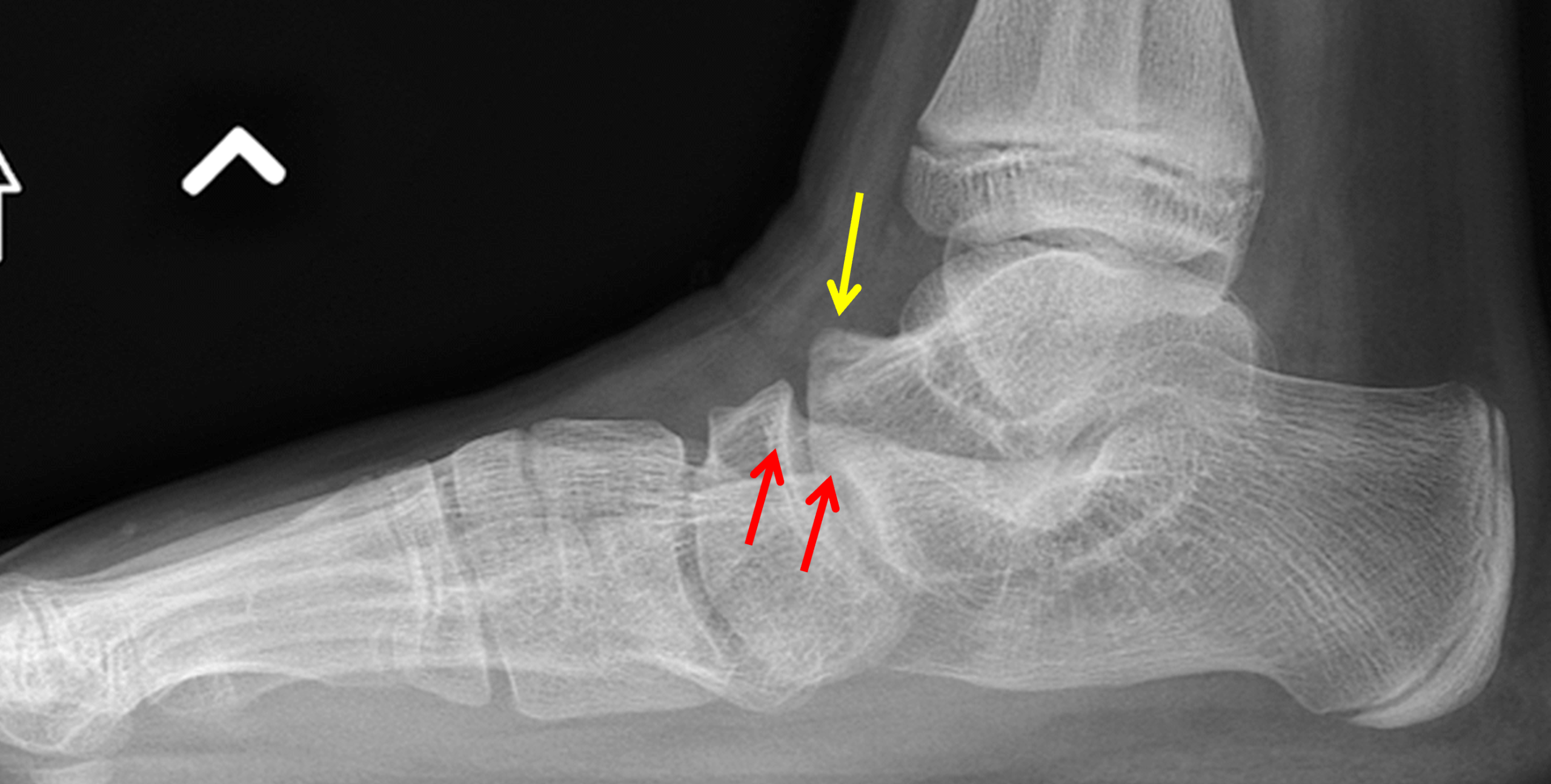

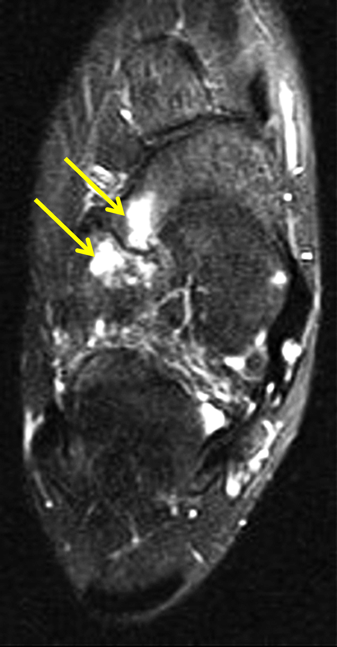
Figure 7: Calcaneonavicular coalition in a 12 year-old. (7A) Lateral radiograph shows mild plantar flexion of the talus and decreased calcaneal pitch. The anterior process of the calcaneus is elongated (red arrows) and a dorsal talar beak is present (yellow arrow). (7B) Long-axis T1-weighted image demonstrates an abnormal articulation between the anterior calcaneus (C) and the lateral aspect of the tarsal navicular bone (N) representing a non-osseous coalition with secondary degenerative changes. There is medial orientation of the long axis of the talus consistent with hindfoot valgus. (7C) Long-axis fat-suppressed, T2-weighted image reveals marrow edema (arrows) in the navicular and calcaneus adjacent to the coalition site.


Figure 8: Talocalcaneal (subtalar) coalition in a 14-year-old girl. (8A) Non-weightbearing lateral radiograph shows a short talar neck and a positive “C-sign” created by the contour of the talar dome and horizontally oriented sustentaculum talus (arrowheads). A hypertrophied dorsal talar beak (arrow) is the most common secondary sign of talocalcaneal coalition and is related to subluxation of the tarsal navicular bone. (8B) T1-weighted sagittal image depicts the fibrous coalition of the middle facets of the talus and calcaneus (yellow arrows). Note the hypertrophied talar beak (red arrow). (8C) CT coronal reconstruction demonstrates fibrous fusion of across the middle subtalar joint (arrow) with altered orientation of the bones and sclerotic changes of the middle talar facet and sustentaculum talus.
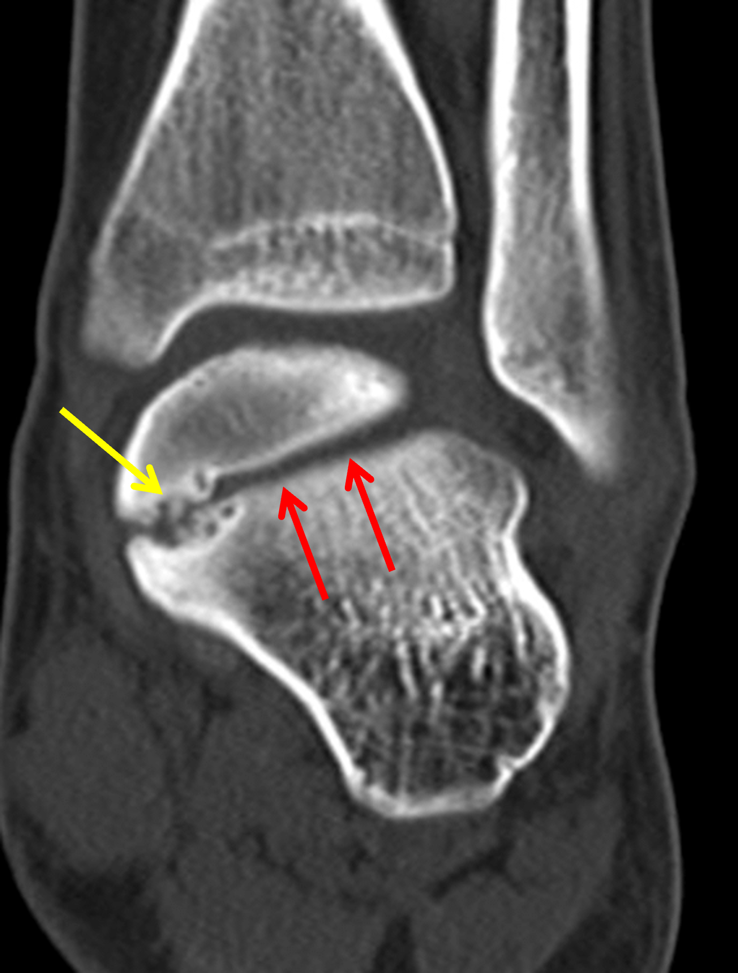

Figure 9: Extra-articular subtalar coalition in a 14 year-old girl. (9A) Coronal CT reconstruction shows a non-osseous coalition (yellow arrow) located medial to the posterior subtalar joint characterized by interdigitation of the facing bone surfaces with degenerative cyst formation and bone sclerosis. Broadening of the lateral process of the talus is a secondary sign of talocalcaneal coalition, possibly related to chronic adaptive changes in response to hindfoot valgus. The posterior subtalar joint (red arrows) has an abnormal medial angulation. (9B) Sagittal fat-suppressed, T2-weighted image shows marrow edema surrounding the extra-articular coalition (arrows), which is located posterior to the middle subtalar joint.
Hindfoot Impingement
An AALTF without an associated coalition can cause painful lateral hindfoot impingement and subfibular impingement, and present with a painful rigid flatfoot and sinus tarsi tenderness.26 The findings of AALTF are subtle on radiographs and are often overlooked (Figure 10A). On a lateral radiograph or sagittal MRI or reconstructed CT, a bisector of the angle of Gissane can be drawn and act as a clue in subtle cases (Figure 2B).27 Associated MRI findings include talocalcaneal marrow edema, sinus tarsi fat abnormalities and dorsal talar beaking (Figure 2). CT may show sclerotic and cystic changes at the site of impingement (Figure 10B). The angle of Gissane is smaller in individuals with an AALTF, with or without associated symptoms.
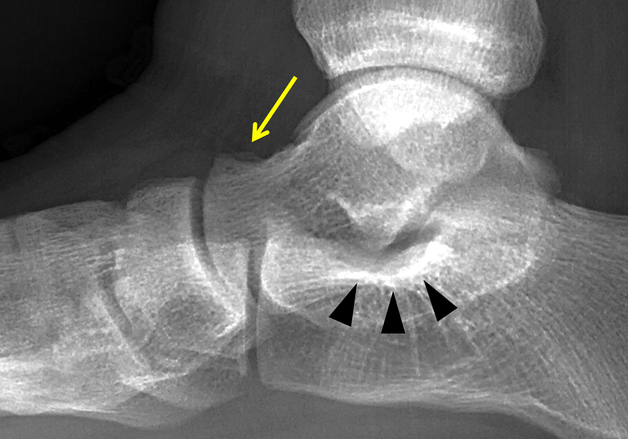

Figure 10: Two symptomatic patients with an AALTF and imaging findings suggesting talocalcaneal impingement. (10A) Lateral radiograph in a 15-year-old boy with painful flatfoot and stiffness shows an AALTF and mild bone sclerosis at angle of Gissane (arrowheads). A small dorsal talar beak is present (arrow). (10B) Sagittal reconstructed CT image in a 14-year-old girl with painful flatfoot and stiffness shows a prominent AALTF (arrow) articulating with the anterior process of the calcaneus. Note the mild sclerotic bone changes at the angle of Gissane (arrowheads). A portion of a dorsal talar beak is partially visualized. An associated talocalcaneal coalition was also present (not shown).
In flatfoot with severe hindfoot valgus, the lateral margin of the calcaneus approaches the lateral malleolus, which may cause symptoms due to subfibular impingement (Figure 11). MRI findings include marrow edema and cyst formation, thickening and edema of the regional ligaments, soft tissue ganglion formation, and diffuse fibrotic changes and soft tissue edema at the inferior fibular tip.

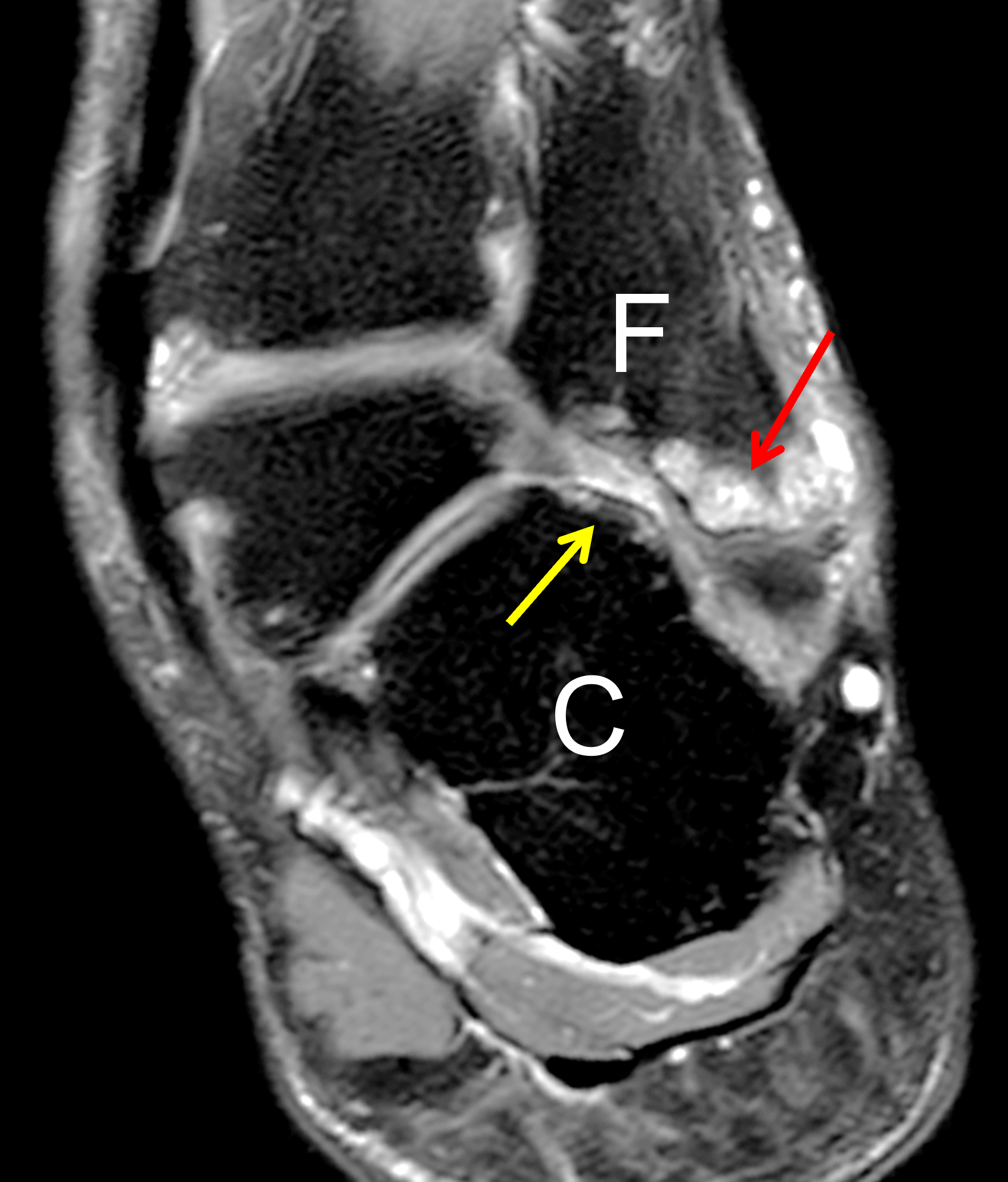
Figure 11: Subfibular impingement in a patient with a history of a previously corrected clubfoot deformity. (11A) Mortise projection radiograph shows hindfoot valgus and degenerative cysts in the inferomedial aspect of the lateral malleolus (arrow). Note the arthritis involving the lateral aspect of the tibiotalar joint. (11B) Coronal fat-suppressed, fluid-sensitive image shows severe hindfoot valgus with the superolateral calcaneus (C) abutting the inferior aspect of the distal fibula (F). Extensive cyst formation is present in the inferior fibula (red arrow) as well as mild marrow edema in the adjacent calcaneus (yellow arrow) and synovitis between the two bones.
Cerebral Palsy
Different types of chronic foot deformity are frequently seen in patients with cerebral palsy. Flatfoot is the most common deformity in children with bilateral lower extremity spasticity.13 Hindfoot valgus is typically present in combination with forefoot abduction and hallux valgus (Figure 12).


Figure 12: Foot malalignments in a 16-year-old girl with cerebral palsy. (12A) Weightbearing lateral radiograph shows plantar flexion of the talus and decreased calcaneal pitch, representing pes planus. (12B) Weightbearing AP (dorsoplantar) view demonstrates severe hindfoot valgus, uncovering of the talar head, and forefoot abduction with hallux valgus.
Overcorrection of clubfoot
Surgical release related to correction of clubfoot may result in a complex flatfoot with the hindfoot in valgus (Figure 11) and dorsal subluxation of the navicular bone (Figure13).28 Clinically, there is often a prominent dorsal bunion, caused by dorsal subluxation of the navicular bone and a tilted first metatarsal.
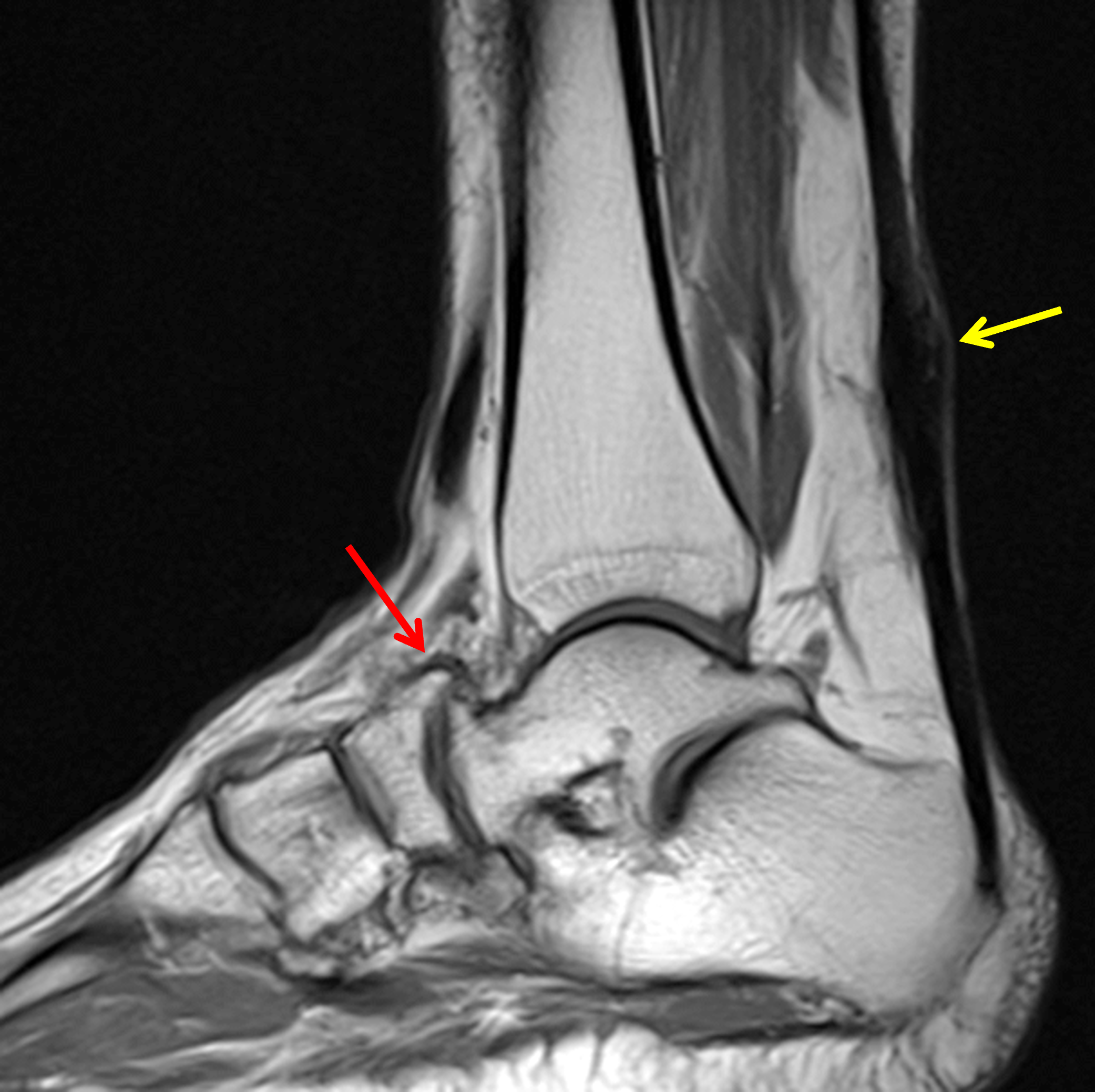

Figure 13: Hindfoot malalignment in an adult who underwent extensive surgical release in childhood for correction of clubfoot deformity. (13A) Sagittal T1-weighted image shows decreased calcaneal pitch. Note the dorsal subluxation and secondary arthritis at the talonavicular joint (red arrow). Focal thickening of the Achilles tendon (yellow arrow) is related to a tendon lengthening procedure. (13B) Coronal fat-suppressed, T2-weighted image shows hindfoot valgus. The deformed calcaneus (C) is shifted laterally.
Painful Type II Accessory Navicular
The accessory navicular bone can be a sesamoid in the distal posterior tibial tendon (Type I) or an accessory ossicle into which the posterior tibial tendon inserts (Type II). The Type II accessory navicular bone articulates with the medial aspect of the tarsal navicular through a synchondrosis. While most cases are asymptomatic, pain can develop if there is trauma from a direct blow, overuse or chronic irritation related to shoe wear. A symptomatic accessory navicular may demonstrate marrow edema in the accessory bone or navicular, and/or high signal intensity in the synchondrosis (Figure 14). Further details concerning the MRI findings are covered in a prior Radsource MRI Web Clinic. There is an increased incidence of painful accessory navicular syndrome in children and adults with flexible flatfoot deformity, presumably caused by direct pressure from the talus on the synchondrosis and tensile forces related to the posterior tibial tendon.20
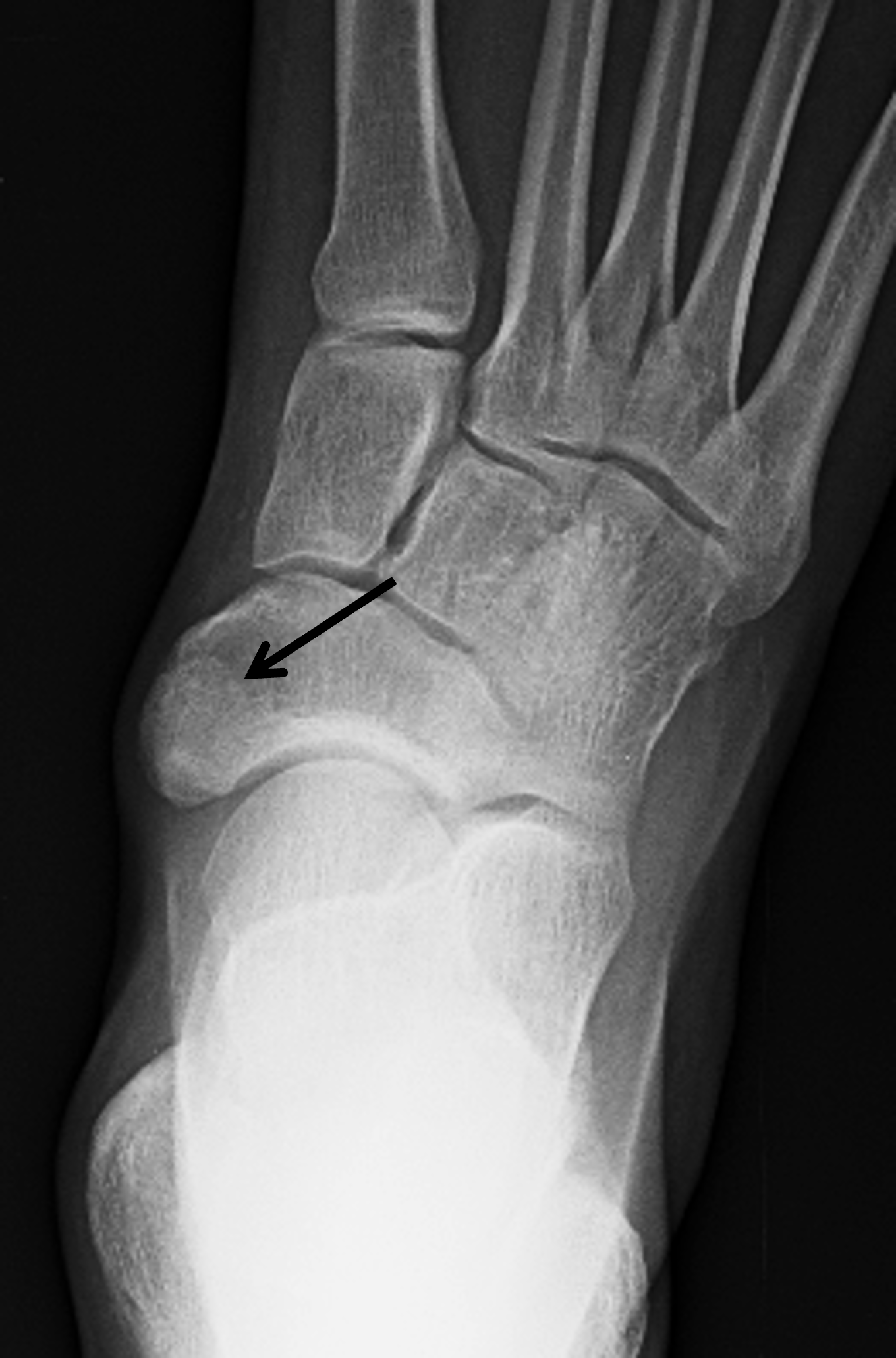
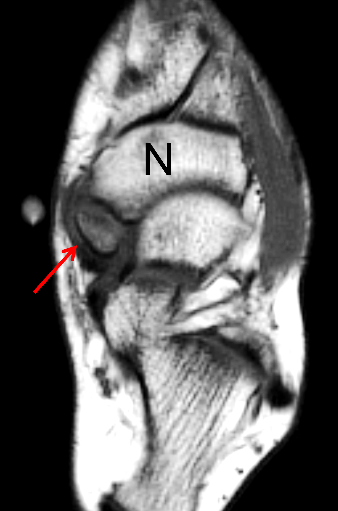
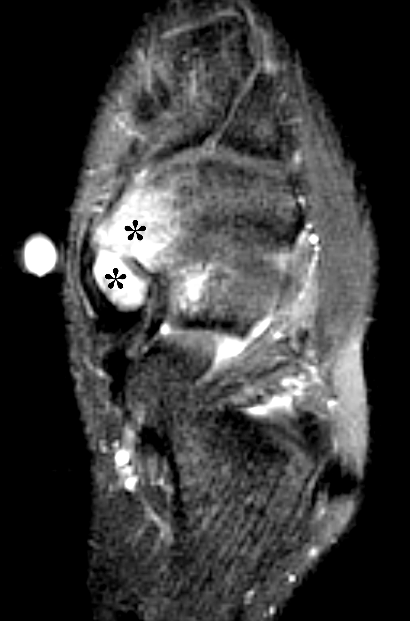
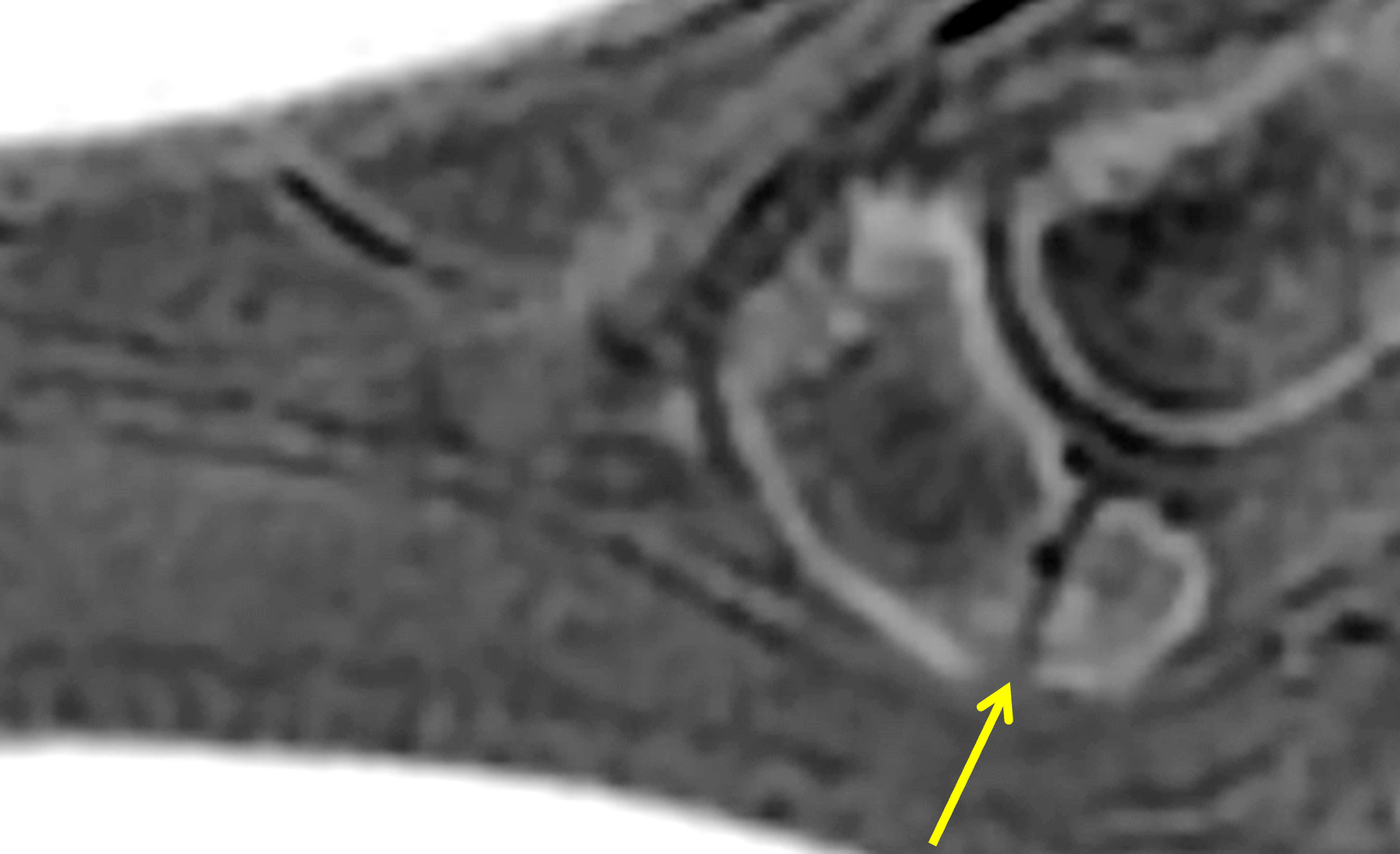
Figure 14: Painful accessory navicular syndrome in two adolescents with flatfoot. (14A) Dorsoplantar radiograph in a 16-year-old girl shows a Type II accessory ossicle (arrow) superimposed on the navicular bone. Mild hindfoot valgus is present. (14B) Corresponding long-axis T1-weighted image shows the accessory bone (arrow) articulating with the medial aspect of the tarsal navicular (N). (14C) Long-axis fat-suppressed, fluid-sensitive image demonstrates extensive marrow edema in the accessory ossicle and the medial aspect of the navicular (asterisks). Symptoms resolved after surgical resection of the ossicle and reimplantation of the posterior tibial tendon (Kidner procedure). (14D) In a second 17-year-old girl, a sagittal MR image obtained with ZTE (zero echo time) technique creates a CT-like image showing cortical irregularity, mild sclerosis and small osseous spurs at the anterior margin of the synchondrosis of a Type II accessory navicular bone (arrow), consistent with chronic microinstability at the synchondrosis.
Stress Fractures and Physeal Stress Injury in Pediatric Flatfoot
There is an increased risk for stress fractures in pediatric flatfeet caused by the altered biomechanics and reduced shock absorption (Figure 15). Fractures have been observed in the navicular, metatarsals (most commonly the second or third), calcaneus and distal fibula.29 Findings on radiographs may be subtle or absent and some patient may need CT or MRI for further evaluation.
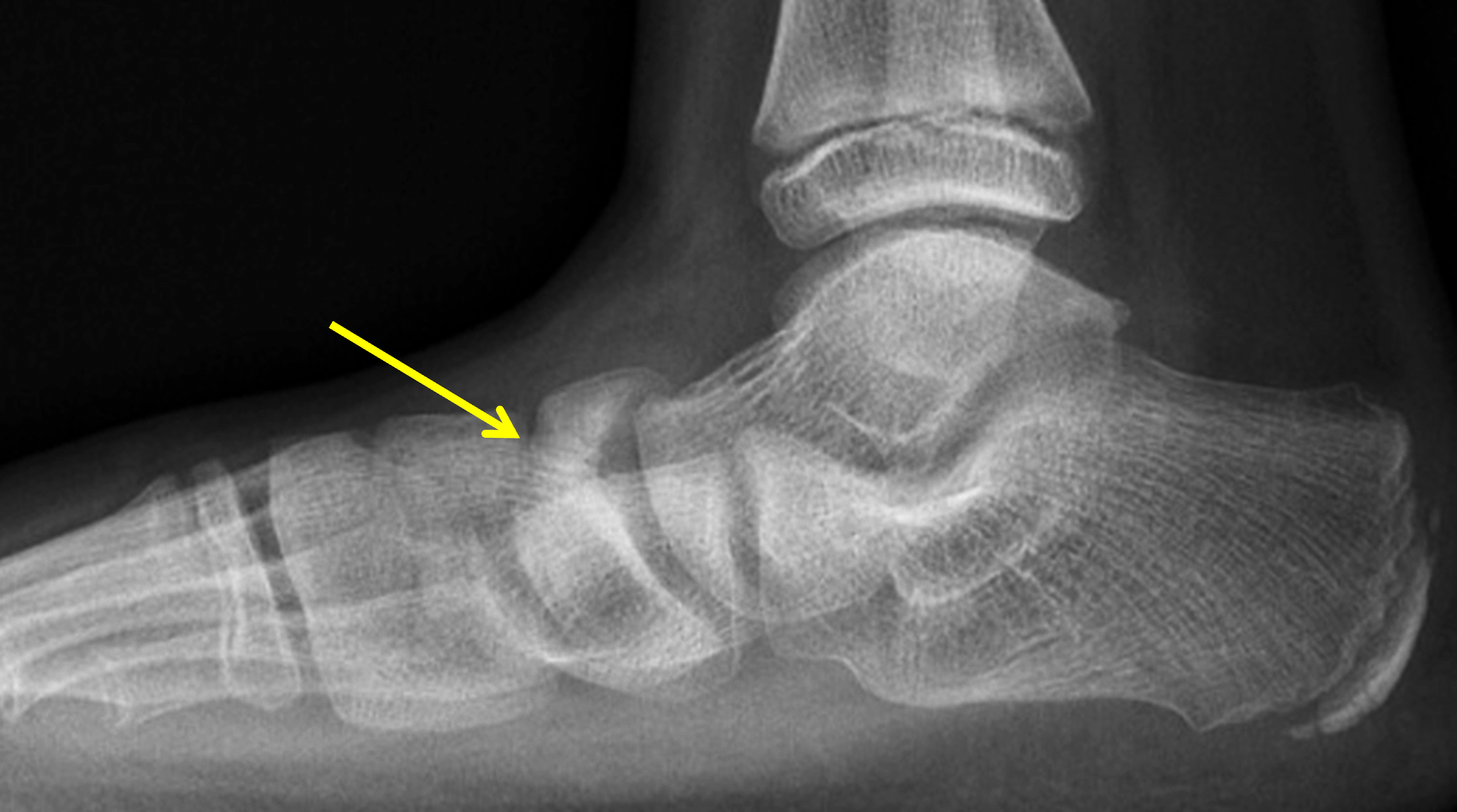


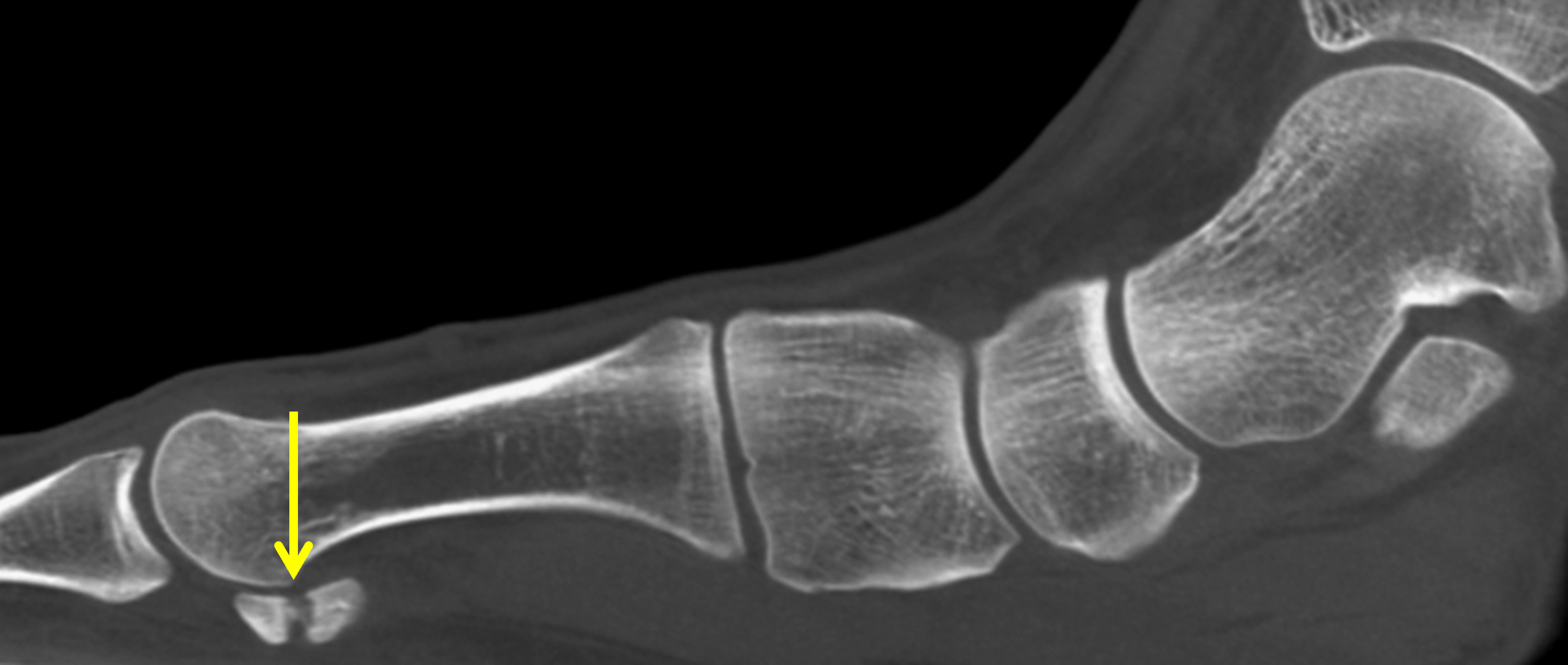
Figure 15: Examples of stress fractures in pediatric patients with flatfoot deformity. (15A) Lateral weightbearing radiograph in an 8-year-old girl shows sclerosis and collapse of the tarsal navicular (arrow) representing a stress fracture. Prompt diagnosis is important because limited blood supply to the central navicular may delay healing. (15B) AP (dorsoplantar) radiograph in an 11-year-old boy demonstrates healing stress fractures of the second metatarsal base and first metatarsal metaphysis characterized by linear sclerosis representing intraosseous callus (arrows). Pronation of the foot related to flatfoot deformity leads to abnormal load on the proximal metatarsals. (15C) T1-weighted sagittal image in a 9-year-old boy with a history of flatfoot and medial mid foot pain. Radiographs were reported as negative. Mild physeal widening in the first metatarsal base and irregularity of the physeal margins (arrow) are signs of stress injury. (15D) Sagittal CT image in a 15-year-old girl with chronic medial forefoot pain. A healing fracture involves the medial hallux sesamoid (arrow). Overpronation associated with flatfoot leads to abnormal forces that increase the risk of sesamoid fractures at the first metatarsal head. The talus is plantar flexed.
Flat foot is associated with overpronation, which leads to increased tension on the Achilles tendon and at its insertion on the calcaneal apophysis. The posterior calcaneal physis is the weak link of the muscle-tendon-bone chain, which responds with osseous remodeling of the adjacent apophysis after chronic exposure to traction and shearing forces. The resulting clinical condition – considered a traction apophysitis – is also known as Sever disease.30 It is a common cause of heel pain in individuals aged 8 to 15 years, with a more common occurrence in males, and is usually diagnosed based on clinical evaluation. A short or tight Achilles tendon is thought to be a contributing factor. Radiographs are usually not necessary for diagnosis and are often normal but may show widening and stress-related changes in the posterior calcaneal growth plate (Figure 16). Contralateral radiographs may be helpful to differentiate osseous abnormalities from normal variants.

Figure 16: 11-year-old boy with flatfoot deformity and heel pain, diagnosed clinically with Sever disease. Weightbearing lateral radiograph shows widening of the posterior calcaneal growth plate with irregular margins of the adjacent metaphysis (arrows). Plantar flexion of the talus and decreased calcaneal pitch indicate underlying flatfoot deformity.
Management
Treatment decisions should be guided primarily by symptoms, functional limitations, flexibility, and the presence of structural abnormalities. Asymptomatic flexible flatfoot generally requires reassurance and observation rather than active intervention. For symptomatic patients, initial treatment includes supportive footwear, orthoses, physical therapy, and activity modification. Stretching exercises are also helpful in the setting of a tight Achilles tendon and calf muscles. Orthotics will often decrease discomfort and pain, but have no lasting effect upon the shape of the longitudinal arch.31
Surgical treatment is generally reserved for children and adolescents with persistent pain, progressive deformity, or significant functional impairment despite conservative management.32,33,34 The choice of surgical procedure depends on the severity and flexibility of the deformity, skeletal maturity, muscle-tendon contractures, and underlying pathology. Surgery aims to restore appropriate hindfoot alignment, improve talonavicular coverage, reconstruct the medial longitudinal arch, and correct forefoot abnormalities. Most symptomatic flexible flatfoot deformities requiring surgery are treated with a combination of bony realignment and soft tissue procedures.
Surgical reconstruction of flexible flatfoot includes lateral column lengthening, commonly performed through an Evans-type calcaneal osteotomy (Figure 17A), which corrects forefoot abduction and improves talonavicular alignment. A medial displacement calcaneal osteotomy may be used to correct hindfoot valgus (Figure 17B). Gastrocnemius contracture or Achilles tendon shortening may have to be corrected by tenotomy. Persistent forefoot supination after hindfoot correction may be addressed through a medial cuneiform osteotomy (Cotton procedure, Figure 17C) or a first tarsometatarsal joint (Lapidus) arthrodesis (Figure 17D). Subtalar arthroereisis (Figure 18), which involves placement of an implant within the sinus tarsi to restrict excessive subtalar eversion, remains controversial because of variable outcomes and implant related complications. Arthrodesis (fusion) is generally avoided in skeletally immature patients with flexible flatfoot, and is reserved for patients with severe rigid deformity, degenerative changes, or complex neuromuscular conditions.
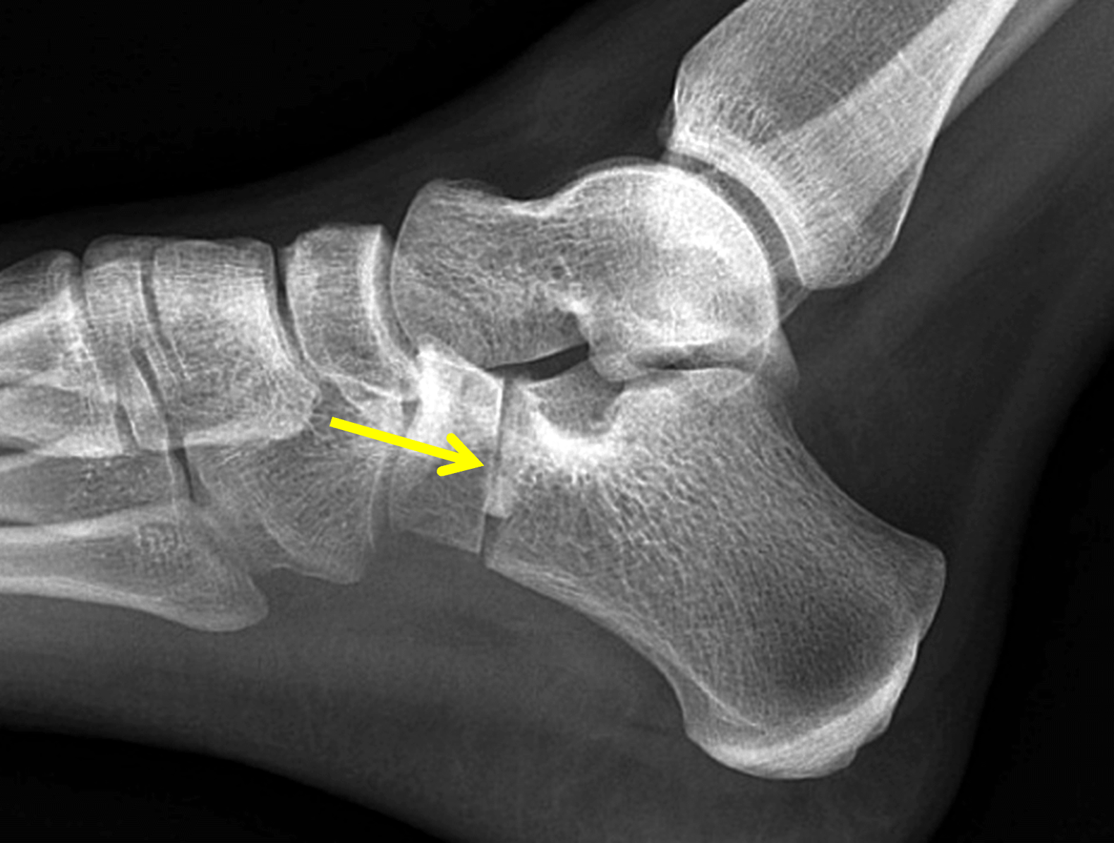

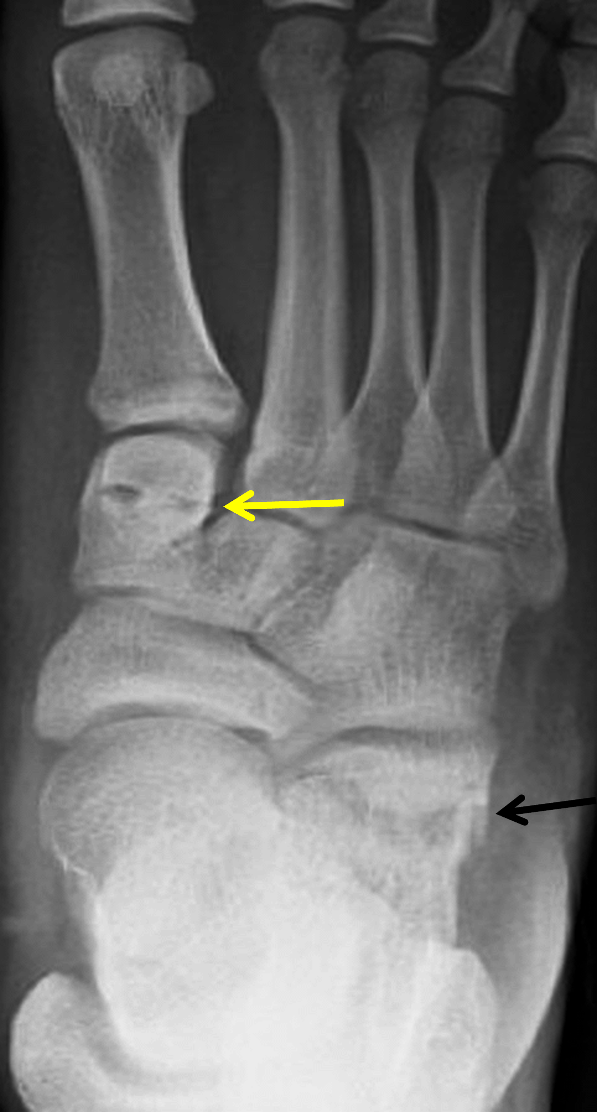

Figure 17: Examples of osteotomies and fusions used to surgically treat flatfoot. (17A) Non-weightbearing lateral radiograph shows a recent wedge (Evans) osteotomy (arrow) in the anterior calcaneus. An interposed bone graft lengthens the lateral column and restores the medial longitudinal arch. (17B) Weightbearing lateral radiograph shows ongoing healing of a medial displacement calcaneal osteotomy (arrow) transfixed with a cannulated screw. The osteotomy realigns the pull of the Achilles tendon and supports the hindfoot. (17C) AP (dorsoplantar) radiograph shows a lateral lengthening calcaneal osteotomy (black arrow) and a medial cuneiform (Cotton) osteotomy (yellow arrow). A Cotton osteotomy addresses residual forefoot varus or supination, allowing the first ray to properly contact the ground. (17D) AP (dorsoplantar) radiograph shows a healed lateral column lengthening osteotomy (black arrow) and an internally-fixated first tarsometatarsal joint (Lapidus) arthrodesis (yellow arrow) to stabilize and restore the medial column of the foot.


Figure 18: Subtalar arthroereisis. (18A) Post-operative lateral radiograph obtained through a fiberglass cast in a 10-year-old with a symptomatic flexible flatfoot, who had pain refractory to conservative measures. An arthroereisis implant has been placed (arrow). A lateral calcaneal osteotomy with an interposition bone graft is transfixed by a pin. (18B) Sagittal T1-weighted image in a different (adult) patient demonstrates a subtalar arthroereisis implant (arrow) located in the sinus tarsi. The implants can be metal or silastic, and act to restrict excessive subtalar joint pronation while elevating the talus. Figure 18A courtesy of Bill Morrison, MD, Philadelphia.
Symptomatic tarsal coalition is managed surgically through coalition resection, sometimes combined with interposition grafting and corrective osteotomy when severe hindfoot valgus is present.35 Arthrodesis may be necessary if the coalition is large or subtalar arthrosis has developed (Figure 19).

Figure 19: Hindfoot fusion for subtalar arthrosis after resection of a talocalcaneal coalition. A non-weightbearing lateral radiograph shows internally-fixated talonavicular and subtalar arthrodeses.
A painful accessory navicular bone associated with flatfoot almost always requires surgical treatment. Complete removal of the accessory ossicle is necessary, followed by reattachment of the posterior tibial tendon (Kidner procedure). Additional procedures may be required to correct pes planus.36
Congenital vertical talus requires early treatment with serial manipulation and casting, followed by reduction and stabilization of the talonavicular joint, with Achilles tenotomy or lengthening as indicated.37
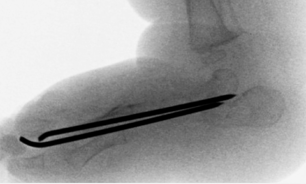
Figure 20: Surgical treatment of congenital vertical talus. Intraoperative lateral fluoroscopic view shows pins used to maintain the position of the talus and navicular after manipulation. The orientation of the talus has been restored.
Conclusion
Most children with flatfoot are asymptomatic and need no further assessment or treatment. Imaging is reserved for symptomatic, rigid, or atypical deformities. Weightbearing radiographs provide information about foot alignment, while MRI and CT are valuable tools when structural abnormalities are suspected. Radiologists can contribute substantially by understanding the normal foot anatomy and alignment in children, and by identifying structural abnormalities. A comprehensive evaluation of the patient that includes not only the imaging results, but also symptoms, flexibility and function, is essential for proper treatment of pediatric flatfoot.
References
- Rose GK, Welton EA, Marshall T. The diagnosis of flat foot in the child. J Bone Joint Surg Br. 1985;67(1):71–78. doi:10.1302/0301-620X.67B1.3968149 ↩
- Harris RI, Beath T. Army foot survey: an investigation of foot ailments in Canadian soldiers. National Research Council Canada; 1952. doi:10.4224/21273359 ↩
- Vanderwilde R, Staheli LT, Chew DE, Malagon V. Measurements on radiographs of the foot in normal infants and children. J Bone Joint Surg Am. 1988;70(3):407–415. ↩
- Metcalfe SA, Bowling FL, Baltzopoulos V, Maganaris C, Reeves ND. The reliability of measurements taken from radiographs in the assessment of paediatric flat foot deformity. Foot (Edinb). 2012;22(3):156–162. doi:10.1016/j.foot.2012.02.006 ↩
- Mosca VS. Flexible flatfoot in children and adolescents. J Child Orthop. 2010;4(2):107–121. doi:10.1007/s11832-010-0239-9 ↩
- Berkeley R, Tennant S, Saifuddin A. Multimodality imaging of the paediatric flatfoot. Skeletal Radiol. 2021;50(11):2133–2149. doi:10.1007/s00256-021-03806-8 ↩
- Staheli LT, Chew DE, Corbett M. The longitudinal arch. A survey of eight hundred and eighty-two feet in normal children and adults. J Bone Joint Surg Am. 1987;69(3):426–428. ↩
- Gould N, Moreland M, Alvarez R, Trevino S, Fenwick J. Development of the child’s arch. Foot Ankle. 1989;9(5):241–245. doi:10.1177/107110078900900506 ↩
- Pfeiffer M, Kotz R, Ledl T, Hauser G, Sluga M. Prevalence of flat foot in preschool-aged children. Pediatrics. 2006;118(2):634–639. doi:10.1542/peds.2005-2126 ↩
- Harris RI, Beath T. Hypermobile flat-foot with short tendo achillis. J Bone Joint Surg Am. 1948;30A(1):116–140. ↩
- Concolino D, Pasquzzi A, Capalbo G, Sinopoli S, Strisciuglio P. Early detection of podiatric anomalies in children with Down syndrome. Acta Paediatr. 2006;95(1):17–20. doi:10.1080/08035250500325108 ↩
- Cass AD, Camasta CA. A review of tarsal coalition and pes planovalgus: clinical examination, diagnostic imaging, and surgical planning. J Foot Ankle Surg. 2010;49(3):274–293. doi:10.1053/j.jfas.2010.02.003 ↩
- Sees JP, Miller F. Overview of foot deformity management in children with cerebral palsy. J Child Orthop. 2013;7(5):373–377. doi:10.1007/s11832-013-0509-4 ↩
- Shuen V, Prem H. Acquired unilateral pes planus in a child caused by a ruptured plantar calcaneonavicular (spring) ligament. J Pediatr Orthop B. 2009;18(3):129–130. doi:10.1097/BPB.0b013e32832b07ca ↩
- Vulcano E, Maccario C, Myerson MS. How to approach the pediatric flatfoot. World J Orthop. 2016;7(1):1–7. doi:10.5312/wjo.v7.i1.1 ↩
- Harris RI, Beath T. Etiology of peroneal spastic flat foot. J Bone Joint Surg Br. 1948;30B(4):624–634. ↩
- Thapa MM, Pruthi S, Chew FS. Radiographic assessment of pediatric foot alignment: review. AJR Am J Roentgenol. 2010;194(6 Suppl):S51–58. doi:10.2214/AJR.07.7143 ↩
- Flores DV, Mejia Gomez C, Fernandez Hernando M, Davis MA, Pathria MN. Adult Acquired Flatfoot Deformity: Anatomy, Biomechanics, Staging, and Imaging Findings. Radiographics. 2019;39(5):1437–1460. doi:10.1148/rg.2019190046 ↩
- Bagley C, McIlhone S, Singla N, Berkeley R, O’Donnell P, Tennant S, et al. MRI for paediatric flatfoot: is it necessary? Br J Radiol. 2022;95(1132):20210784. doi:10.1259/bjr.20210784 ↩
- Kamel SI, Belair JA, Hegazi TM, Halpern EJ, Desai V, Morrison WB, et al. Painful type II os naviculare: introduction of a standardized, reproducible classification system. Skeletal Radiol. 2020;49(12):1977–1985. doi:10.1007/s00256-020-03503-y ↩
- Sandberg JK, Young VA, Yuan J, Hargreaves BA, Wishah F, Vasanawala SS. Zero echo time pediatric musculoskeletal magnetic resonance imaging: initial experience. Pediatr Radiol. 2021;51(13):2549–2560. doi:10.1007/s00247-021-05125-5 ↩
- David MS, Weinraub GM. Calcaneovalgus and Congenital Vertical Talus. In: Butterworth ML, Marcoux JT, editors. The Pediatric Foot and Ankle: Diagnosis and Management. Cham: Springer International Publishing; 2020. p. 163–168. ↩
- Miller M, Dobbs MB. Congenital Vertical Talus: Etiology and Management. J Am Acad Orthop Surg. 2015;23(10):604–611. doi:10.5435/JAAOS-D-14-00034 ↩
- Newman JS, Newberg AH. Congenital tarsal coalition: multimodality evaluation with emphasis on CT and MR imaging. Radiographics. 2000;20(2):321–332; quiz 526–327, 532. doi:10.1148/radiographics.20.2.g00mc03321 ↩
- Alqahtani E, Fliszar E, Resnick DL, Huang BK. Accessory anterolateral talar facet associated with tarsal coalition: prevalence and cross-sectional characterization. Skeletal Radiol. 2020;49(3):417–424. doi:10.1007/s00256-019-03293-y ↩
- Singh JP, Loomba S, Pun B, Virmani S. Non-osseous coalition of accessory anterolateral talar facet-A case series. Skeletal Radiol. 2025;54(4):893–898. doi:10.1007/s00256-024-04781-6 ↩
- Aydıngöz Ü, Topcuoğlu OM, Görmez A, Cankurtaran T, Topcuoğlu ED, Ergen FB. Accessory Anterolateral Talar Facet in Populations With and Without Symptoms: Prevalence and Relevant Associated Ankle MRI Findings. AJR Am J Roentgenol. 2016;207(4):846–851. doi:10.2214/ajr.16.16056 ↩
- Faldini C, Artioli E, Panciera A, Bonelli S, Arceri A, Mazzotti A. (Surgical management of clubfoot overcorrection: a case series). Arch Orthop Trauma Surg. 2023;143(11):6503–6511. doi:10.1007/s00402-023-04946-3 ↩
- Liu J, Granberry W. Pes Planus and Distal FIbular Stress Fractures. Foot & Ankle Orthopaedics. 2017;2(3):2473011417S2473000265. doi:10.1177/2473011417s000265 ↩
- Hendrix CL. Calcaneal apophysitis (Sever disease). Clin Podiatr Med Surg. 2005;22(1):55–62, vi. doi:10.1016/j.cpm.2004.08.011 ↩
- Wenger DR, Mauldin D, Speck G, Morgan D, Lieber RL. Corrective shoes and inserts as treatment for flexible flatfoot in infants and children. J Bone Joint Surg Am. 1989;71(6):800–810. ↩
- Bouchard M, Mosca VS. Flatfoot deformity in children and adolescents: surgical indications and management. J Am Acad Orthop Surg. 2014;22(10):623–632. doi:10.5435/JAAOS-22-10-623 ↩
- Kwon JY, Myerson MS. Management of the flexible flat foot in the child: a focus on the use of osteotomies for correction. Foot Ankle Clin. 2010;15(2):309–322. doi:10.1016/j.fcl.2010.02.001 ↩
- Smith C, Zaidi R, Bhamra J, Bridgens A, Wek C, Kokkinakis M. Subtalar arthroereisis for the treatment of the symptomatic paediatric flexible pes planus: a systematic review. EFORT Open Rev. 2021;6(2):118–129. doi:10.1302/2058-5241.6.200076 ↩
- Kothari A, Masquijo J. Surgical treatment of tarsal coalitions in children and adolescents. EFORT Open Rev. 2020;5(2):80–89. doi:10.1302/2058-5241.5.180106 ↩
- Samsonov AP, Butler JJ, Konar K, Rubin J, Lai Fat-Fur R, Rampersad C, et al. Operative Management for Symptomatic Accessory Navicular: A Systematic Review. Foot Ankle Spec. 202519386400251363022. doi:10.1177/19386400251363022 ↩
- Yang JS, Dobbs MB. Treatment of Congenital Vertical Talus: Comparison of Minimally Invasive and Extensive Soft-Tissue Release Procedures at Minimum Five-Year Follow-up. J Bone Joint Surg Am. 2015;97(16):1354–1365. doi:10.2106/JBJS.N.01002 ↩
