
Clinical History
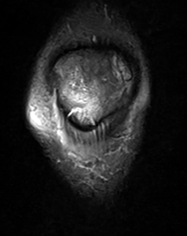
An 11-year-old boy sustained a knee injury while running cross country 2 weeks prior to imaging. Pain is centered at the inferior patella when extending the knee, with associated anterior bruising. (1A) Sagittal fat-suppressed, fluid sensitive, (1B) sagittal proton density-weighted, and (1C) coronal fat-suppressed, fluid-sensitive images are provided. What are the findings? What is your diagnosis?


Findings
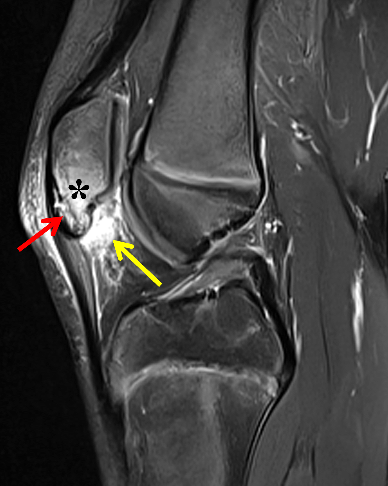

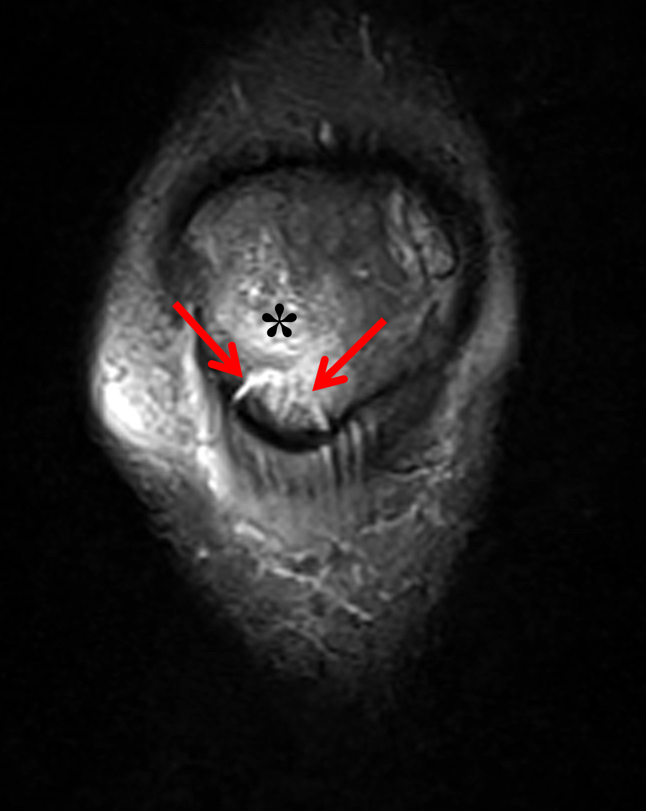
Figure 2: (2A) Sagittal fat-suppressed, fluid-sensitive image shows a non-displaced fracture line (red arrow) through the inferior pole of the patella involving the trabecular bone and and growth cartilage, with marrow edema (asterisk) in the adjacent patella. The proximal patellar tendon is thick and edematous. Soft tissue edema is present in the superior aspect of Hoffa’s fat pad (yellow arrow). (2B) Sagittal proton density-weighted image confirms the fracture (arrow) through the inferior patella. (2C) Coronal fat-suppressed, fluid-sensitive sequence demonstrates the fracture line (arrows) containing a thin cleft of high signal involving the peripheral low-signal rim of growth cartilage and the trabecular bone, with adjacent marrow edema (asterisk). The bones are skeletally immature with open growth plates.
Diagnosis
Inferior patellar sleeve avulsion fracture.
Introduction
Patellar sleeve avulsion fractures are rare injuries of the skeletally immature knee in which a “sleeve” of cartilage together with a variable amount of bone is pulled off the patella – most commonly the inferior pole – by forceful eccentric quadriceps contraction against the resisting patellar tendon.1 The injuries are clinically important because they are easily missed on radiographs yet, when displaced, disrupt the extensor mechanism and require prompt surgical repair.
Anatomy and Pathophysiology
The patella is the largest sesamoid bone in the body, embedded within the extensor mechanism and functioning as an anatomic lever arm that increases the mechanical advantage of the quadriceps pull. It is a hemispherical structure whose deep surface bears a hyaline cartilage lining for articulation with the femoral trochlea. Developmentally, the patella forms not inside the tendon but from the femur via progenitor cells expressing both chondrogenic and tenogenic markers, later separating from the femur by a joint formed under mechanical influence.2
Appreciating the ossification process of the patella is key to understanding the vulnerability of the patella to sleeve avulsion injuries. In the infant and young child, the patella is entirely cartilaginous and radiographically invisible (though detectable on ultrasound and MRI). Ossification begins between 3 and 7 years, usually from a single (77%) but sometimes multiple primary centers that enlarge and coalesce; around age 12, secondary ossification centers may appear, especially superolaterally. Full expansion of the ossified bone to the cartilaginous contours is not complete until late adolescence, coinciding with maturation of the surrounding epiphyseal centers.2
During the window between the onset of ossification and skeletal maturity, a radiolucent cartilage shell surrounds the ossified core (Figure 3A). Initially, the cartilage is relatively thick compared to the central bone nucleus. As skeletal maturity progresses, a process of chondro-osseous transformation occurs, similar to the process of secondary ossification in an epiphysis.3 Over time the ossified portion becomes larger while the peripheral cartilage thins. The enthesis between the patellar tendon and inferior patellar cartilage is mechanically weaker than in the adult where the cartilage is woven into the Sharpey fibers, so forceful quadriceps contraction can avulse a sleeve of cartilage at the inferior patella in children (Figures 3B and 3C).4 The avulsed fragment will contain radiographically-occult cartilage inferiorly with or without a portion of the calcified deepest cartilage layer and underlying trabecular bone. Because the avulsed fragment contains unmineralized cartilage, it is larger than the fragment visible on radiographs, which is often just a small calcified fleck.5
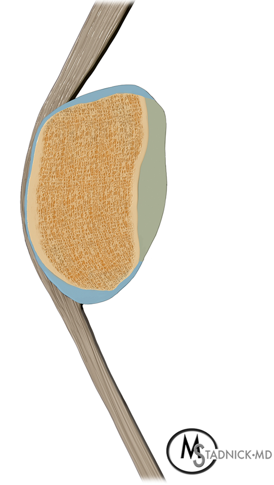
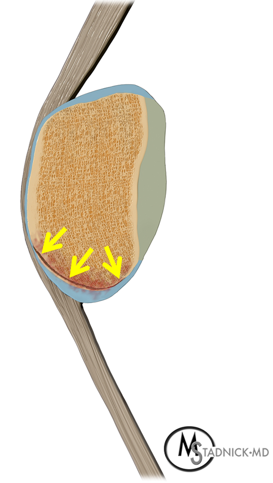
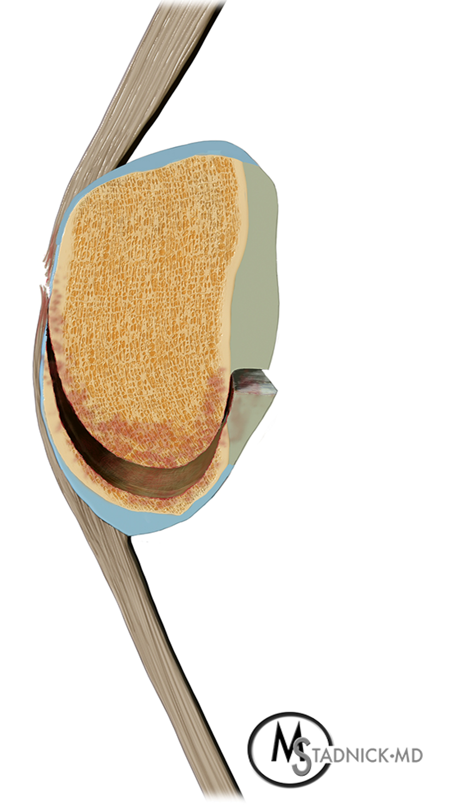
Figure 3. Anatomic basis for inferior patellar sleeve avulsions shown by sagittal cross-sectional illustrations through the patella. The drawings approximate the degree of skeletal maturity in a 12-year-old boy. (3A) The intact patella has a central ossified nucleus surrounded by a layer of growth cartilage (light blue) and hyaline cartilage (light gray). The deepest (yellow) layer of the cartilage is mineralized, representing ongoing chondro-osseous transformation of the cartilage into mature bone. (3B) With a non-displaced sleeve avulsion, the fracture plane is located within the cartilage layer (arrows), resulting in an inferior fragment composed of cartilage with or without a small sliver of bone. The cartilaginous portion of the fragment will be invisible radiographically. (3C) Displaced sleeve avulsions occur when the fracture plane involves the entire thickness of the cartilage layer together with portions of the surrounding extensor mechanism (pre-patellar quadriceps expansion and retinacula).
The inferior (distal) patellar pole is the classic injury site. In an anatomic study of 100 cadaveric specimens, the inferior pole shape was pointed in 57% of knees, intermediate-shaped in 21%, and blunted in 22%. The patellar tendon attachment was classified as either anterior or posterior type, with the posterior type associated with a significantly shorter tendon. These morphologic variants influence local tensile stress distribution, with pointed patellar morphology and anterior tendon attachments impinging during knee flexion due to the posterior tilt of the patella, possibly contributing to sleeve injuries. Conversely, with posterior patellar tendon attachments and blunted inferior patellar shape, strong tensile stress in the posterior tendon fibers may predispose to sleeve avulsion fractures.6
Patellar fractures are rare overall in children, comprising less than 1–2% of pediatric fractures, but sleeve fractures account for the majority (roughly 57–72%) of patellar fractures in this age group.7 Injuries predominate in boys, peaking in the pre-adolescent to adolescent years; reported cohorts have mean ages ranging from about 10–15 years, with most injuries sport-related or traumatic.8 The typical mechanism is a noncontact injury with sudden forceful quadriceps contraction, as occurs when landing from a jump.9 Less commonly the injury occurs from blunt force direct trauma like in a dashboard injury.7
Clinically, patients present with focal pain and tenderness at the inferior pole of the patella. Associated soft tissue swelling and ecchymoses are common. On physical examination, the patella may be high riding, with a palpable gap along its inferior surface. If the fracture extends intra-articularly, a knee effusion will be present. Patients may not be able to actively extend the knee against gravity or bear weight without pain.4,10
Typical Imaging Findings and Pitfalls
Especially for nondisplaced injuries, diagnosis requires a high index of suspicion based on mechanism, effusion, and extensor lag, because the avulsed fragment is largely radiolucent cartilage.11 Lateral radiographs may show a small osseous fragment at the inferior pole and unilateral patella alta (Figure 4), which is an important clue – sleeve fractures show significantly higher Insall-Salvati ratios (mean ~1.9) compared to other disorders involving the inferior pole.12 Avulsion fractures may displace at the time of injury or in a delayed fashion if the initial injury is missed and the patient continues activities and sports. Once displaced, the pull of the quadriceps can result in a large fracture gap and severe patella alta (Figure 5). Displaced avulsions require operative reduction and fixation to restore extensor mechanism function. In very young children, the only radiographic findings of a sleeve avulsion may be soft tissue swelling at the inferior pole of the patella with or without patella alta.3,9
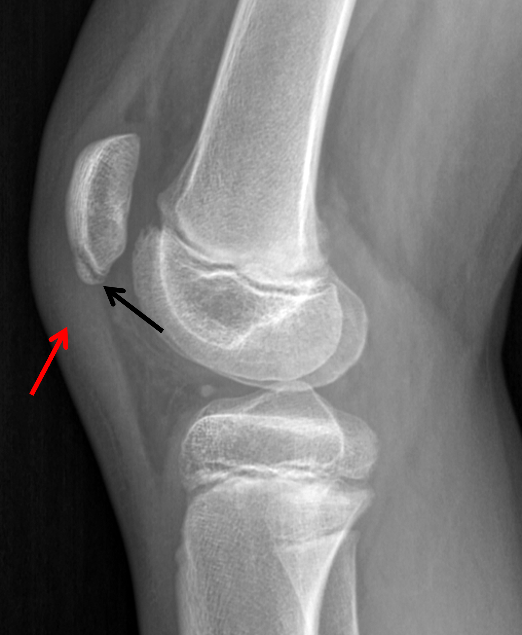
Figure 4: Inferior patellar sleeve avulsion in an 11- year-old boy after acute trauma. Lateral radiograph shows a nondisplaced avulsed bone fragment at the inferior pole of the patella (black arrow) representing the mineralized portion of a larger fragment comprised of bone and unossified cartilage. The adjacent proximal patellar tendon appears thick (red arrow) with an indistinct anterior margin. Patella alta is present (Insall-Salvati ratio = 1.5; Caton-Deschamps ratio = 1.6).


Figure 5: Displaced inferior patellar sleeve avulsion in a 12-year-old boy. The initial injury was weeks before and radiographs (not available) were reported as normal. He continued playing sports and developed sudden increased pain and inability to extend the knee. (5A) Lateral radiograph shows a complete patellar sleeve avulsion with severe inferior displacement of the fracture fragment (arrow), a wide fracture gap, severe patella alta, and anterior soft tissue swelling. (5B) Subsequent lateral acquired six weeks after open reduction and internal suture fixation demonstrates the reduced, healed fracture resulting in an elongated contour of the inferior patella (arrow), similar to the appearance of healed Sinding-Larsen-Johansson syndrome.
On radiographs, several pitfalls should be kept in mind. First, accessory secondary ossification centers can mimic a fracture. While the most common location for an accessory ossification center is the superolateral aspect of the patella (resulting in a bipartite patella if there is incomplete incorporation of the secondary center after skeletal maturity), an inferior accessory ossification center is also relatively common.13 These are typically thin, curved bone fragments occurring along the anteroinferior margin of the patella and may be bilateral (Figure 6B), although inferior sleeve fractures can also occur bilaterally.14 Sometimes the secondary ossification center may appear slightly displaced from the remainder of the patella, further mimicking a sleeve injury.15 Adding to confusion is the observation that these secondary inferior ossification centers are reported more frequently in athletic children,3 and also commonly appear in children with spasticity like in cerebral palsy,16 which has led investigators to conclude that at least some of them are actually unrecognized prior avulsion fractures.3,15
Sinding-Larsen-Johansson (SLJ) syndrome is a chronic painful condition in adolescents, felt to be analogous to Osgood-Schlatter disease. In SLJ syndrome, focal tenderness and swelling are present at the inferior pole of the patella, accompanied by mature ossification in the proximal patellar tendon.15 Chronic or healed SLJ lesions may result in an elongated appearance of the inferior patella, similar to a healed, displaced patellar sleeve fracture (Figure 5B). Like Osgood-Schlatter condition, SLJ syndrome is believed to be traumatic and related to overuse, with the soft tissue ossification either representing metaplasia within the tendon or an united prior avulsion fracture. In an individual that is currently asymptomatic, it may be impossible to distinguish an inferior secondary ossification center on a lateral radiograph from a prior sleeve avulsion fracture or SLJ syndrome. In patients with an acute injury and symptoms, a sleeve avulsion fracture is most likely. If the presentation is with subacute or chronic inferior patellar pain, a greater amount of anterior soft tissue swelling, a larger joint effusion, larger bone fragments, and greater degrees of fragment displacement favor a patellar sleeve injury over SLJ syndrome.12
MRI or ultrasound are useful when the injury is suspected despite normal or equivocal radiographs (Figure 6).4,11,14 MRI is also frequently advocated in acute injuries even when the diagnosis is certain based on clinical presentation and radiographs, because radiographs underestimate the size of the fracture fragment (showing only the ossified portion) and are unable to show the degree of displacement when the injury is entirely cartilaginous, both of which affect the choice of treatment. On MRI, marrow edema will be present in the inferior patella on fluid-sensitive sequences.5 A fracture line through the growth cartilage often appears higher in signal intensity compared to the cartilage on fluid-sensitive sequences (Figures 2C and 6C), unlike typical fracture lines within bone that are usually low signal (Figure 2A). The radiologist should note the size of the fracture fragment (including its cartilaginous portion), the degree of inferior displacement/retraction, extension to the articular surface, the amount of patella alta, and the integrity of the patellar tendon. Involvement of the patellar retinacula should also be noted, because a torn retinaculum will need to be repaired at the time of surgery to preserve full knee extension.7
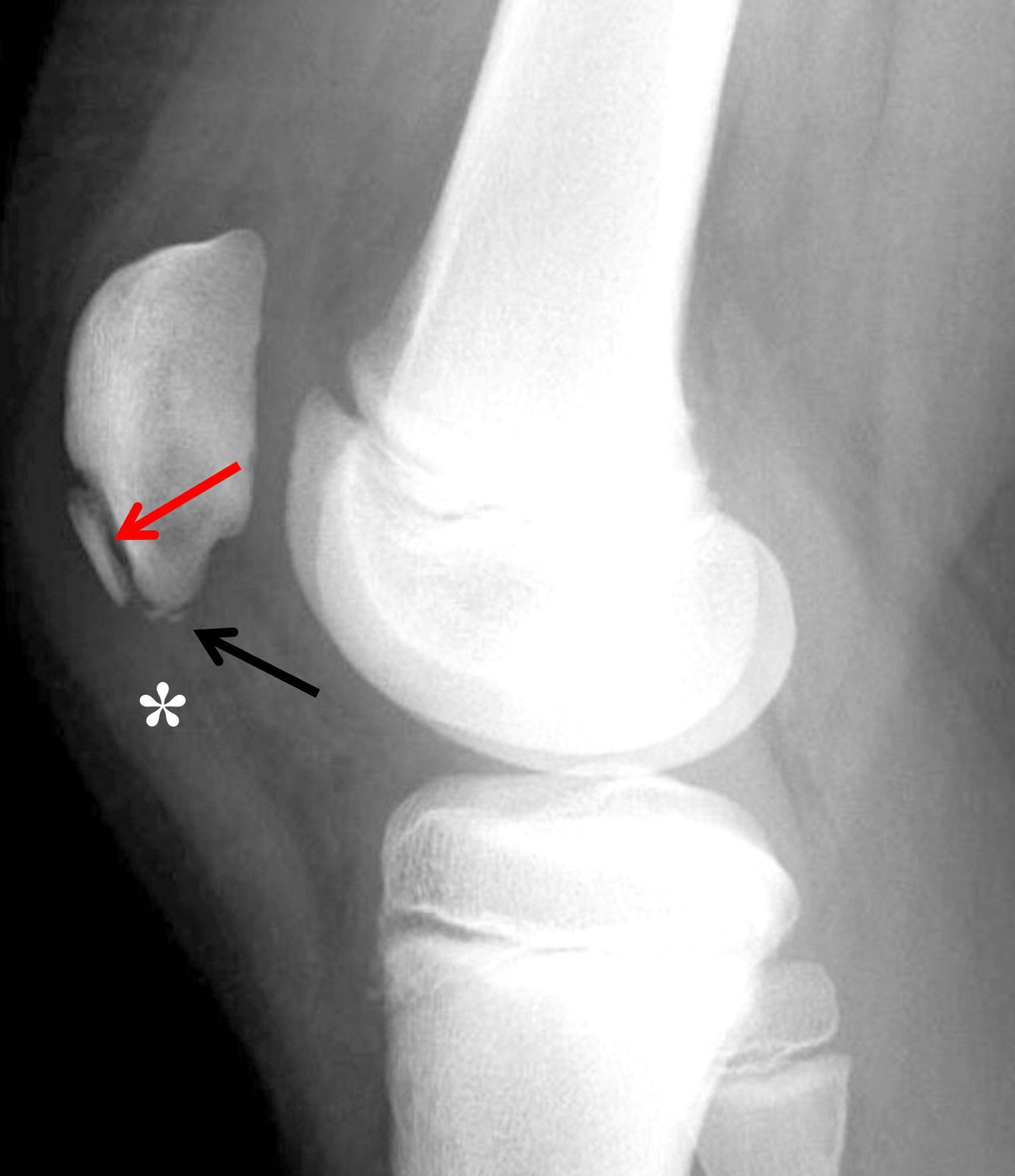

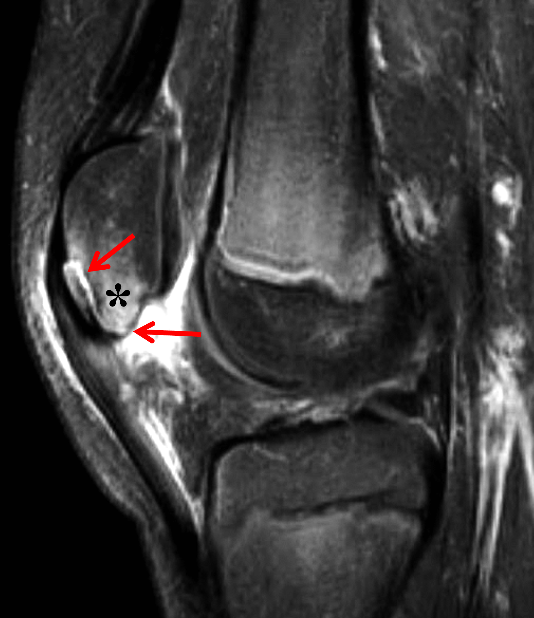
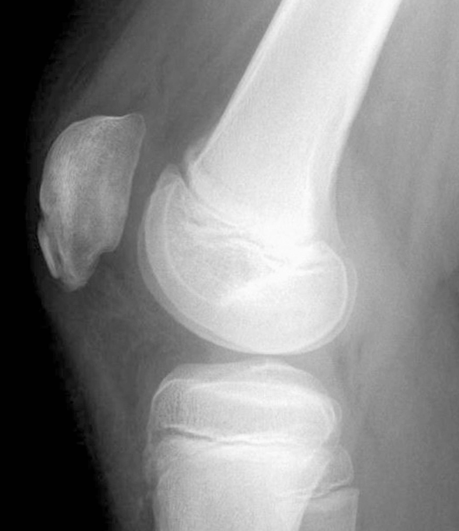
Figure 6: Inferior patellar sleeve avulsion fracture. (6A) Lateral radiograph of the symptomatic side shows a curved separate bone fragment along the anteroinferior patellar margin (red arrow) and two small calcifications inferior to the patella (black arrow). The proximal patellar tendon is thickened (asterisk) and edema is present in Hoffa’s fat pad. (6B) Lateral radiograph of the asymptomatic contralateral knee shows a similar-appearing ossified fragment along the anteroinferior patella (red arrow), though with a smaller gap with the underlying patella compared to 6A. The lack of symptoms and soft tissue changes suggests an accessory ossification center rather than a fracture. (6C) Sagittal fat-suppressed, fluid sensitive image of the symptomatic side demonstrates that the anterior and inferior bone fragments on the radiograph are both contained within a large fragment of bone and cartilage separated from the patella by a high signal fracture line (red arrows). Marrow edema is present in the inferior patella (asterisk) as well as increased signal and thickening of the proximal patellar tendon, and soft tissue edema superficial to the patellar tendon and in Hoffa’s fat pad. (6D) Lateral radiograph a few months later shows partial healing of the anteroinferior and inferior bone fragments.
Variants of Sleeve Injuries
In addition to the inferior patella, similar sleeve injuries can occur less frequently at other locations in the extensor mechanism. Avulsions of the superior patellar growth cartilage, with or without an osseous fragment, have been reported at the quadriceps tendon insertion site (Figure 7).17,18 Additionally, sleeve fractures along the medial patella can accompany lateral patellar dislocation injuries in children.3 And injuries at the distal patellar tendon insertion can avulse a variable-size sleeve of tissue in the proximal tibia.19 Unlike the more common tibial tubercle avulsion fractures, a proximal tibial sleeve injury will also strip off a portion of the proximal tibial periosteum inferior to the growth plate (Figure 8). Rare instances of patellar sleeve injuries have also been reported in adults, at least involving the superior patella.20,21 Sleeve injuries occurring after skeletal maturity are rare because the adult enthesis is much stronger, so that the same mechanism of injury that produces a sleeve avulsion in children, is much more likely to result in a transverse patellar fracture, or rupture of the patellar tendon or quadriceps tendon when there is underlying tendinosis.
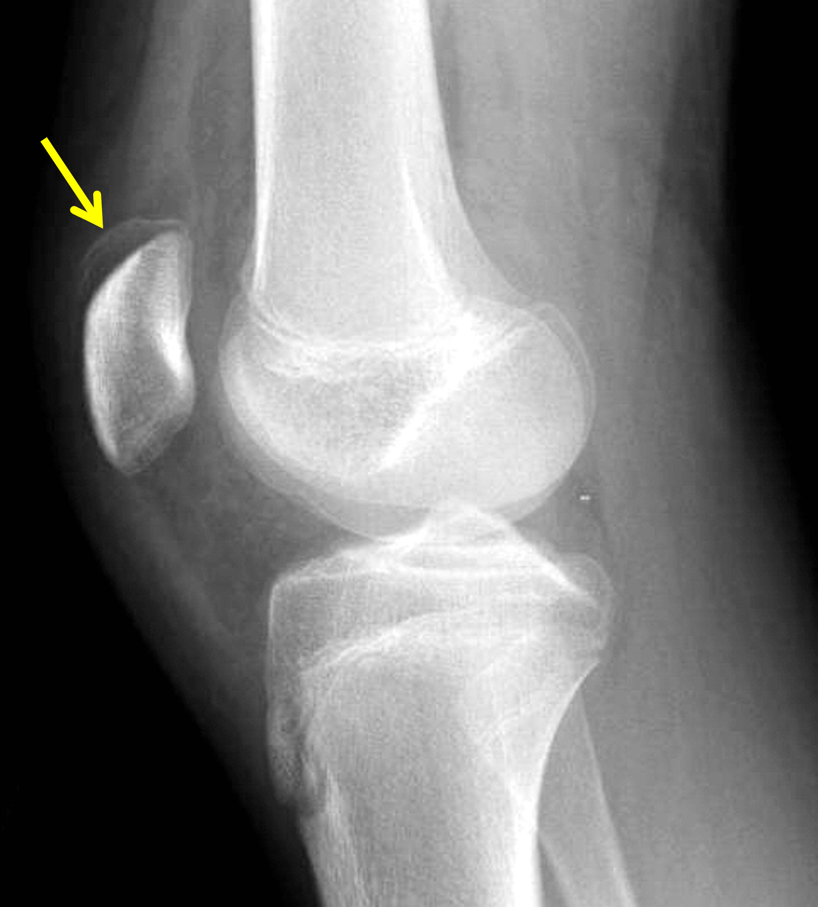
Figure 7: Superior pole patellar sleeve avulsion fracture in an adolescent patient. Lateral radiograph demonstrates a thin shell of bone (arrow) that has been avulsed from the superior patella at the quadriceps tendon insertion.


Figure 8: Proximal tibial sleeve avulsion fracture in a 12-year-old boy due to a sports injury. (8A) Lateral radiograph shows soft tissue swelling anterior to the proximal tibial epiphysis (asterisk) and a thin fracture fragment (arrows) elevated from the proximal tibial metaphysis representing stripped periosteum and underlying bone. Sagittal (8B) fat-suppressed, fluid sensitive and (8C) proton density-weighted images show a partial avulsion of the patellar tendon from the anterior tibial tubercle apophysis (red arrows) and the anterior periosteum and cortex of the tibial metaphysis distal to the growth plate (yellow arrows). Note the marrow edema in the anterior epiphysis (asterisk in 8B).
Differential Diagnosis
Clinically, in addition to inferior sleeve fractures, acute post-traumatic pain at the inferior pole of the patella in a child or adolescent can be related to abnormalities of the inferior patella or surrounding soft tissues. Bone contusions or fracture through the ossified part of the inferior patella are typically due to direct injuries, like from a fall onto the kneecap. Acute osteochondral and chondral lesions of the patella and femoral trochlea also occur. A combination of radiographs and, when necessary, MRI usually suffice to identify infractions of the bone and articular cartilage. Tendinosis and tears of the proximal patellar tendon are usually due to repetitive overuse injuries. Prepatellar bursitis and abnormalities of Hoffa’s fat pad (impingement, contusions) can also produce infrapatellar pain; MRI and ultrasound are the primary imaging methods to identify these soft tissue abnormalities.
As discussed above, the principle radiographic mimics of inferior sleeve avulsions are SLJ syndrome and inferior pole fractures (Figure 9). Clinically, only ~24% patients with SLJ syndrome report acute trauma, and most individuals have preserved straight-leg raising and ability to weight-bear, in contrast to those with acute sleeve fractures. However, overlap can occur, and patellar sleeve avulsion has been reported superimposed on pre-existing SLJ syndrome.11
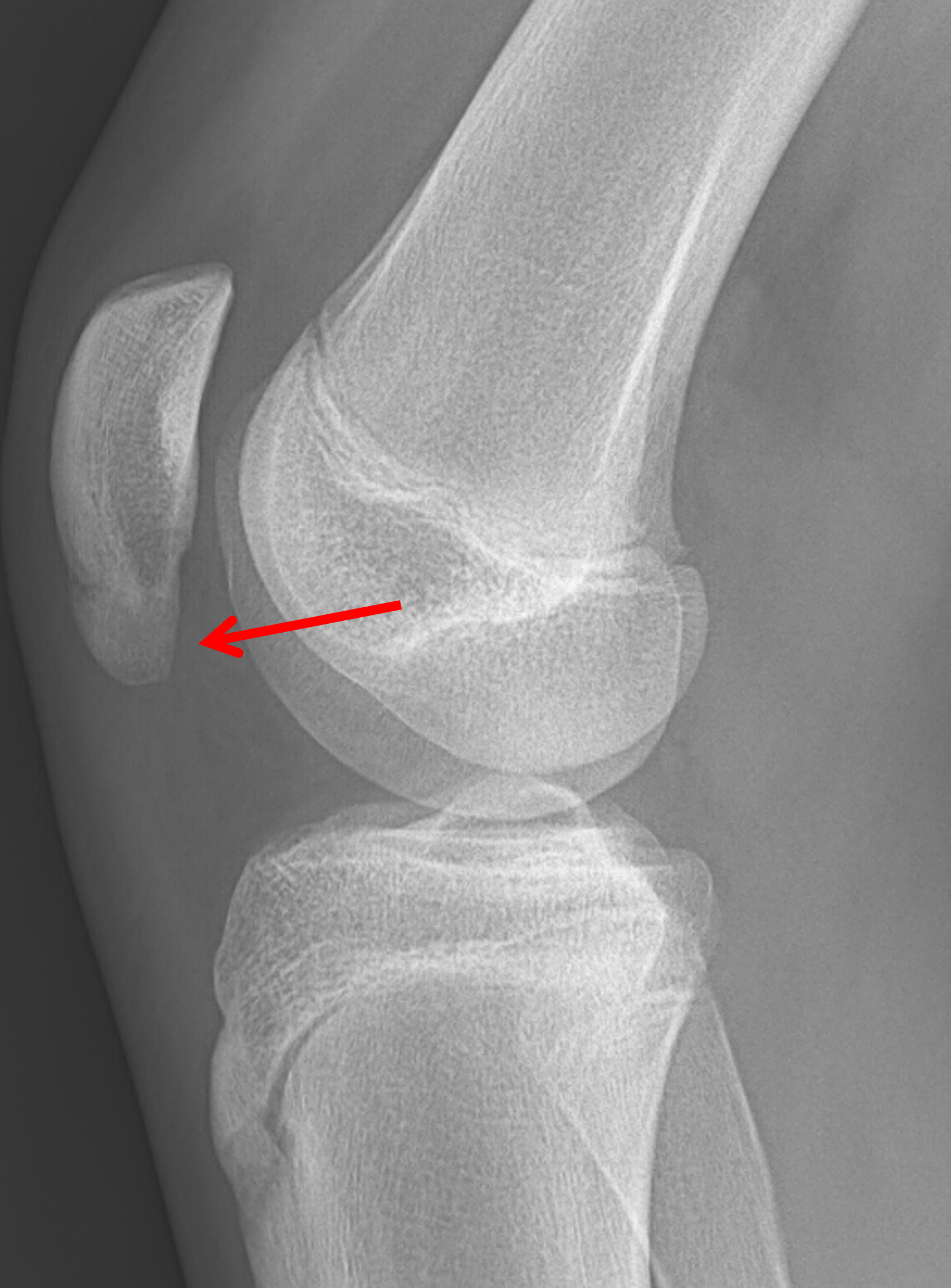
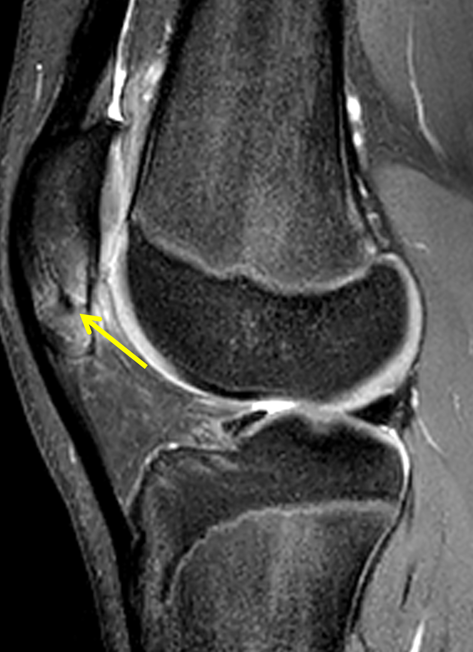
Figure 9: 14-year-old boy with a healed inferior pole patellar fracture and continued pain. (9A) Lateral radiograph shows a healed traction-type fracture through the caudal patella, resulting in an elongated inferior pole (arrow) and patella infera. (9B) Sagittal fat-suppressed, fluid-sensitive image demonstrates the remnant of the fracture line (arrow) and persistent marrow edema representing ongoing inflammation.
Management
Treatment is guided primarily by fragment displacement and extensor mechanism integrity, mirroring general patellar fracture principles where surgery is indicated for loss of extensor mechanism, articular displacement >2 mm, or fragment separation >3 mm. Minimally displaced fractures (typically <2 mm) with an intact extensor mechanism can be managed nonoperatively with immobilization in near-full extension, and case series report excellent healing and functional outcomes.8,22 Displaced fractures require open reduction and internal fixation, restoring the extensor mechanism. Transosseous suture repair (including high-strength suture tape) is a well-described technique with reliably good outcomes; other constructs include tension-band wiring, suture anchors, and newer anchor loop plates.22,23
Delayed or missed diagnosis is associated with worse outcomes. Because the avulsed cartilage retains osteogenic potential, unhealed fragments often continue to form new bone, especially under the continued pull of the extensor mechanism.9 Delayed presenters in one nonoperative cohort ultimately required operative treatment. Reported complications after surgery include patellar tendon ossification, residual extensor lag, loss of flexion, and loose bodies – which may require additional procedures for debridement, loose body removal, or revised fixation – although modern repair techniques report low construct failure rates.8
Conclusion
Patellar sleeve avulsion fractures are uncommon but important pediatric injuries that can be easily overlooked, particularly on radiographs where the largely cartilaginous avulsed fragment may be inconspicuous. Prompt recognition of the characteristic clinical and imaging findings is essential to distinguish these injuries from normal developmental variants and other patellar pathology, as missed or delayed diagnosis may result in extensor mechanism dysfunction, patella alta, and chronic functional impairment. A high index of suspicion, careful evaluation of the patellar tendon and surrounding soft tissues, and appropriate use of MRI or ultrasound when radiographs are equivocal can facilitate timely diagnosis and management.
References
- MacDonald J, Rodenberg R, Sweeney E. Acute Knee Injuries in Children and Adolescents: A Review. JAMA Pediatr. 2021;175(6):624–630. doi:10.1001/jamapediatrics.2020.6130 ↩
- Vanlerberghe C, Boutry N, Petit F. Genetics of patella hypoplasia/agenesis. Clin Genet. 2018;94(1):43–53. doi:10.1111/cge.13209 ↩
- Grogan DP, Carey TP, Leffers D, Ogden JA. Avulsion fractures of the patella. J Pediatr Orthop. 1990;10(6):721–730. doi:10.1097/01241398-199011000-00004 ↩
- Sessions WC, Herring M, Truong WH. Extensor Mechanism Injury in the Pediatric Population-A Clinical Review. J Knee Surg. 2018;31(6):490–497. doi:10.1055/s-0038-1625955 ↩
- Bates DG, Hresko MT, Jaramillo D. Patellar sleeve fracture: demonstration with MR imaging. Radiology. 1994;193(3):825–827. doi:10.1148/radiology.193.3.7972832 ↩
- Edama M, Kageyama I, Nakamura M, Kikumoto T, Nakamura E, Ito W, et al. Anatomical study of the inferior patellar pole and patellar tendon. Scand J Med Sci Sports. 2017;27(12):1681–1687. doi:10.1111/sms.12858 ↩
- Ray JM, Hendrix J. Incidence, mechanism of injury, and treatment of fractures of the patella in children. J Trauma. 1992;32(4):464–467. doi:10.1097/00005373-199204000-00010 ↩
- Sousa PL, Stuart MJ, Prince MR, Dahm DL. Nonoperative Management of Minimally Displaced Patellar Sleeve Fractures. J Knee Surg. 2021;34(3):242–246. doi:10.1055/s-0039-1694742 ↩
- Hunt DM, Somashekar N. A review of sleeve fractures of the patella in children. Knee. 2005;12(1):3–7. doi:10.1016/j.knee.2004.08.002 ↩
- Houghton GR, Ackroyd CE. Sleeve fractures of the patella in children: a report of three cases. J Bone Joint Surg Br. 1979;61-B(2):165–168. doi:10.1302/0301-620X.61B2.438267 ↩
- Schmidt-Hebbel A, Eggers F, Schutte V, Achtnich A, Imhoff AB. Patellar sleeve avulsion fracture in a patient with Sinding-Larsen-Johansson syndrome: a case report. BMC Musculoskelet Disord. 2020;21(1):267. doi:10.1186/s12891-020-03297-z ↩
- Devana SK, Trivellas A, Bennett A, Jackson N, Beck JJ. Clinical and Radiographic Differentiation of Pediatric Patellar Sleeve Fractures and Other Inferior Pole Pathologies. Am J Sports Med. 2022;50(4):977–983. doi:10.1177/03635465221073995 ↩
- Maloney E, Stanescu AL, Ngo AV, Parisi MT, Iyer RS. The Pediatric Patella: Normal Development, Anatomical Variants and Malformations, Stability, Imaging, and Injury Patterns. Semin Musculoskelet Radiol. 2018;22(1):81–94. doi:10.1055/s-0037-1608004 ↩
- Guy SP, Marciniak JL, Tulwa N, Cohen A. Bilateral sleeve fracture of the inferior poles of the patella in a healthy child: case report and review of the literature. Adv Orthop. 2011;2011428614. doi:10.4061/2011/428614 ↩
- Ozonoff MB. Pediatric Orthopedic Radiology, 2nd Ed. Philadelphia: W. B. Saunders; 1992. ↩
- Kaye JJ, Freiberger RH. Fragmentation of the lower pole of the patella in spastic lower extremities. Radiology. 1971;101(1):97–100. doi:10.1148/101.1.97 ↩
- Kimball MJ, Kumar NS, Jakoi AM, Tom JA. Subacute superior patellar pole sleeve fracture. Am J Orthop (Belle Mead NJ). 2014;43(1):29–32. ↩
- Siddiqui R, Singh A, Cullinan C. Superior Pole Sleeve Fracture of the Patella in an Adolescent: A Case Report. JBJS Case Connect. 2020;10(3):e20.00007. doi:10.2106/jbjs.Cc.20.00007 ↩
- Kosuge DD, Balaji VB, Ahad N, Vemulapalli K. Proximal Tibial Sleeve Fracture: Case Report of a Rare Injury and Review of the Literature. Eur J Trauma Emerg Surg. 2010;36(4):388–391. doi:10.1007/s00068-009-9077-1 ↩
- Xie L, Xu H, Zhang L, Xu R, Guo Y. Sleeve fracture of the adult patella: Case report and review of the literature. Medicine (Baltimore). 2017;96(32):e7096. doi:10.1097/md.0000000000007096 ↩
- Kushwaha SS, Kumar N, Bharti A, Maurya G. Neglected Isolated Sleeve Fracture of the Superior Pole of the Patella in a Healthy Adult – A Case Report and Review of Literature. J Orthop Case Rep. 2021;11(1):104–107. doi:10.13107/jocr.2021.v11.i01.1984 ↩
- Gao GX, Mahadev A, Lee EH. Sleeve fracture of the patella in children. J Orthop Surg (Hong Kong). 2008;16(1):43–46. doi:10.1177/230949900801600111 ↩
- Perkins CA, Egger AC, Willimon SC. Transosseous Repair of Patellar Sleeve Fractures: A Case Series and Surgical Technique Guide. J Knee Surg. 2022;35(12):1326–1332. doi:10.1055/s-0041-1723013 ↩
