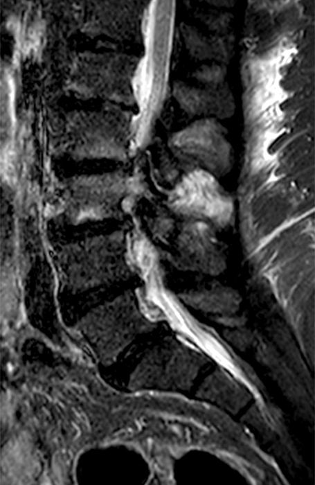
Facet Synovitis
Clinical History: A 69-year-old female presents with low back pain, bilateral buttock, and posterior right thigh pain, aggravated when moving from seated to standing position. The patient denies radicular type symptoms. On exam, the patient demonstrates increased...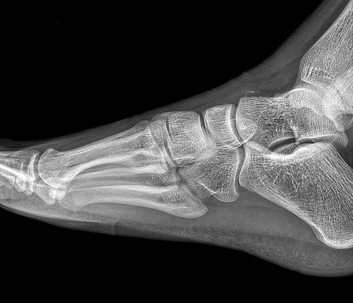
Midtarsal (Chopart) Joint Sprain
Clinical History: A 30 year-old male presents with pain in the midfoot following a twisting injury to the ankle. A lateral radiograph of the foot is provided [Figure 1A] with sagittal fat-suppressed T2-weighted [Figure 1B, 1C and 1D], and axial fat-suppressed...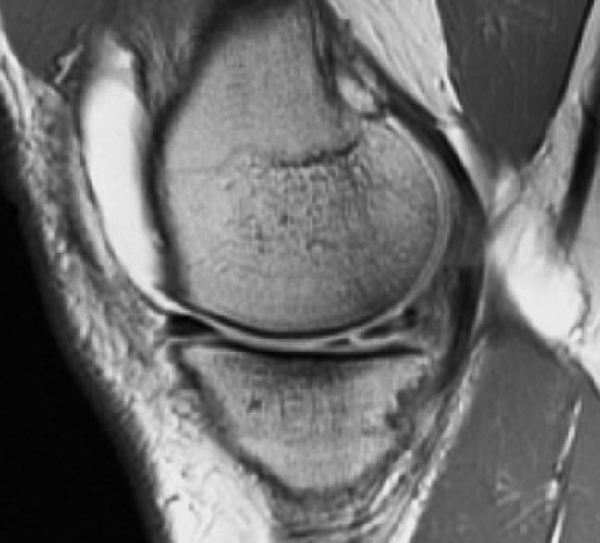
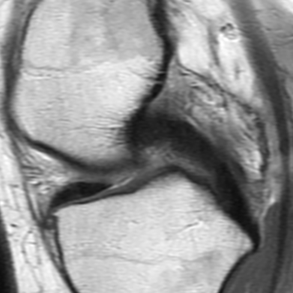
The Postoperative Meniscus
Clinical History: An 18 year-old male with a history of a posterior horn medial meniscus peripheral longitudinal tear treated with meniscal repair at age 16 presents for MR imaging. The patient had a recent new injury with increased pain. (1A) Proton...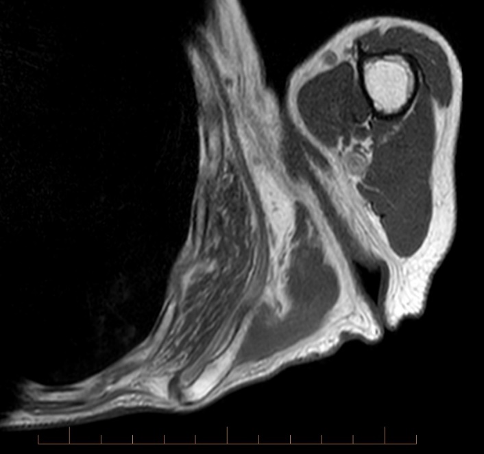
Elastofibroma Dorsi
Clinical History: A 79 year-old female noticed a painless mass along the inferior scapula, present for several months. Axial T1-weighted (1a), coronal T2-weighted (1b), sagittal T1-weighted (1c), and fat-suppressed sagittal T2-weighted (1d) images are provided. What...To provide the highest quality clinical and technology services to customers and patients, in the spirit of continuous improvement and innovation.
Sign up for our monthly web clinicsGet in Touch
Radsource, LLC
750 Old Hickory Blvd, Suite 1-260
Brentwood, TN 37027
ph 615.376.7502
[email protected]


