Radsource radiologists are constantly communicating and sharing knowledge with each other. In our blog series Today’s Interesting Case, our team will post notable cases and images for discussion from time to time.
Today, we are exploring the case of a 62 year-old female presenting after a fall 2 months earlier with shoulder pain and markedly restricted movement.
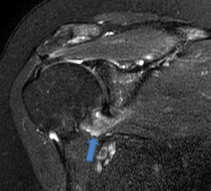
A fat-suppressed T2-weighted coronal image demonstrates marked soft-tissue thickening and edema about the inferior joint capsule.
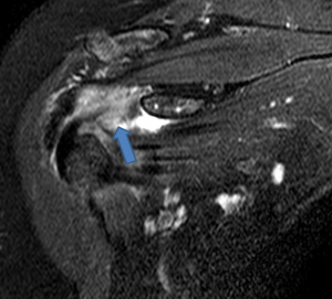
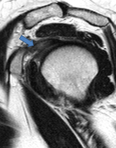
An additional coronal image and a T2-weighted sagittal image also reveal soft-tissue thickening and edema within the rotator interval.
This case is a particularly clear example of the diagnosis of adhesive capsulitis via MR.
As Dr. Stadnick explains in the February 2005 web clinic on the topic, “MRI effectively demonstrates the rotator interval and the axillary recess, which are sites commonly affected by adhesive capsulitis.
“The MRI changes of adhesive capsulitis are most often observed at the rotator interval and the inferior glenohumeral ligament. Recent evidence indicates that IV gadolinium enhanced MR provides even greater specificity in diagnosing adhesive capsulitis. MRI is therefore an invaluable tool in differentiating adhesive capsulitis from other conditions that may have a similar clinical presentation.”