
Clinical History
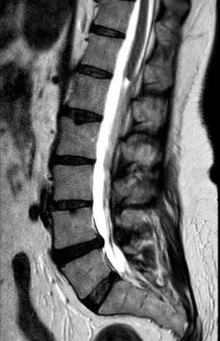
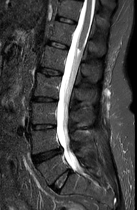
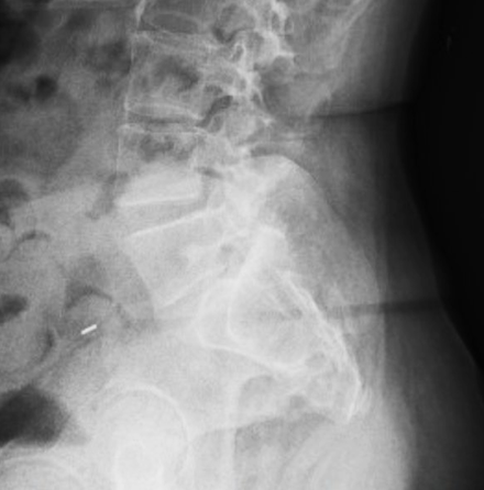
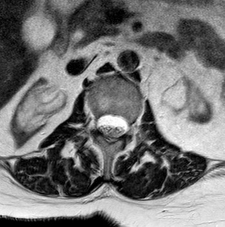
A 57-year-old woman presents with low back pain, neurogenic bladder, and chronic kidney disease. Sagittal (1A) T2-weighted and (1B) STIR images as well as (1C) a lateral radiograph of the lumbar spine are shown. (1D) An additional axial T2-weighted image is provided. What are the findings? What is your diagnosis?
Findings


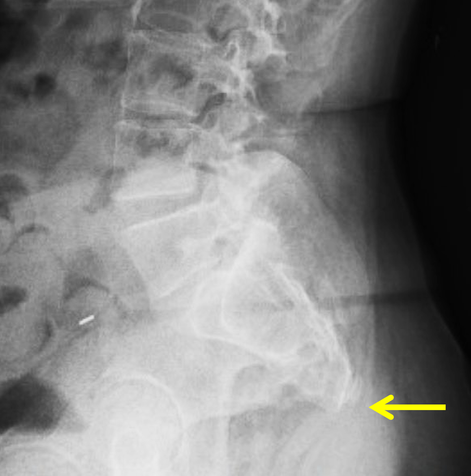
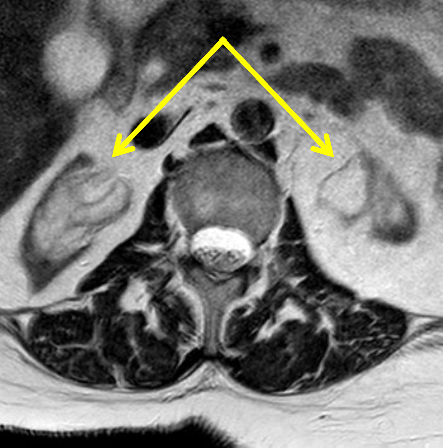
Figure 2: (2A) Sagittal T2-weighted and (2B) Sagittal STIR images of the lumbar spine demonstrate a truncated, blunted, wedge-shaped conus medullaris (arrows) terminating at T12 with a characteristic double-bundle configuration of the proximal cauda equina nerve roots. There is complete absence of the coccyx and partial sacral agenesis (asterisks) with S2 representing the most caudal fully-formed vertebral segment. (2C) Lateral radiograph confirms partial sacral agenesis with absence of the inferior sacrum (arrow) and coccyx. (2D) Axial T2-weighted demonstrates end stage atrophy of both kidneys with dilatation of the renal collecting systems (arrows) representing chronic obstructive uropathy that was due to a neurogenic bladder.
Diagnosis
Caudal Regression Syndrome
Introduction
Caudal regression syndrome (CRS) is a rare congenital disorder characterized by abnormal development of the caudal embryo, resulting in a spectrum of distal spinal abnormalities.1 Spinal involvement ranges from partial coccygeal agenesis to complete lumbosacral agenesis (Figure 3). Given the shared embryologic origin, caudal spinal agenesis is often associated with abnormalities of the genitourinary tract (e.g., renal agenesis or hydronephrosis), gastrointestinal tract (e.g., anorectal malformations and anal atresia) and musculoskeletal abnormalities of the lower extremities such as contractures and clubfoot deformities.2 Coexistent spinal canal and cord abnormalities are also common and include tethered cord, spinal dysraphism, diastematomyelia, terminal hydromyelia, and intraspinal lipomas. More extensive spinal agenesis is generally associated with more severe extra-spinal abnormalities.3
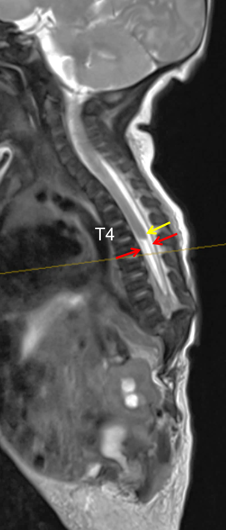

Figure 3: Caudal regression syndrome in a 5-day-old infant with multiple anomalies. (3A) Sagittal T2-weighted image reveals complete lumbosacral agenesis as well as absence of any vertebral structures caudal to T9. The spinal cord terminates at T4 and has a blunted appearance, with the dorsal margin more caudal than the ventral margin (yellow arrow). The distal nerve roots are divided into anterior and posterior bundles (red arrows). (3B) Axial T2-weighted image at the T5-T6 level shows an absent distal cord with the nerve roots located peripherally (arrow). Images courtesy of Franz J. Wippold, M.D.
Imaging is central to the diagnosis and characterization of CRS. Prenatal ultrasound may suggest the diagnosis by demonstrating abnormal sacral development (Figure 4), which can be confirmed and further characterized with fetal MRI. However, many cases are not recognized until after birth. Postnatal radiographs and CT delineate the extent of vertebral agenesis. MRI provides critical information regarding spinal cord morphology, conus termination, and associated spinal abnormalities.
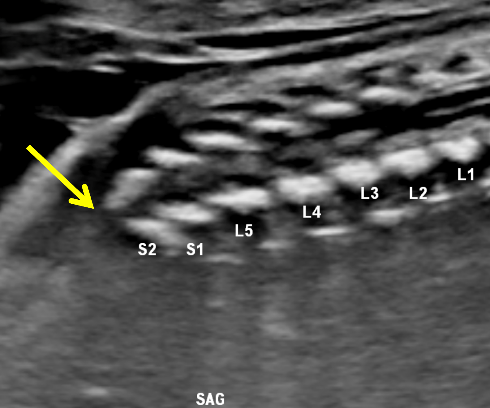
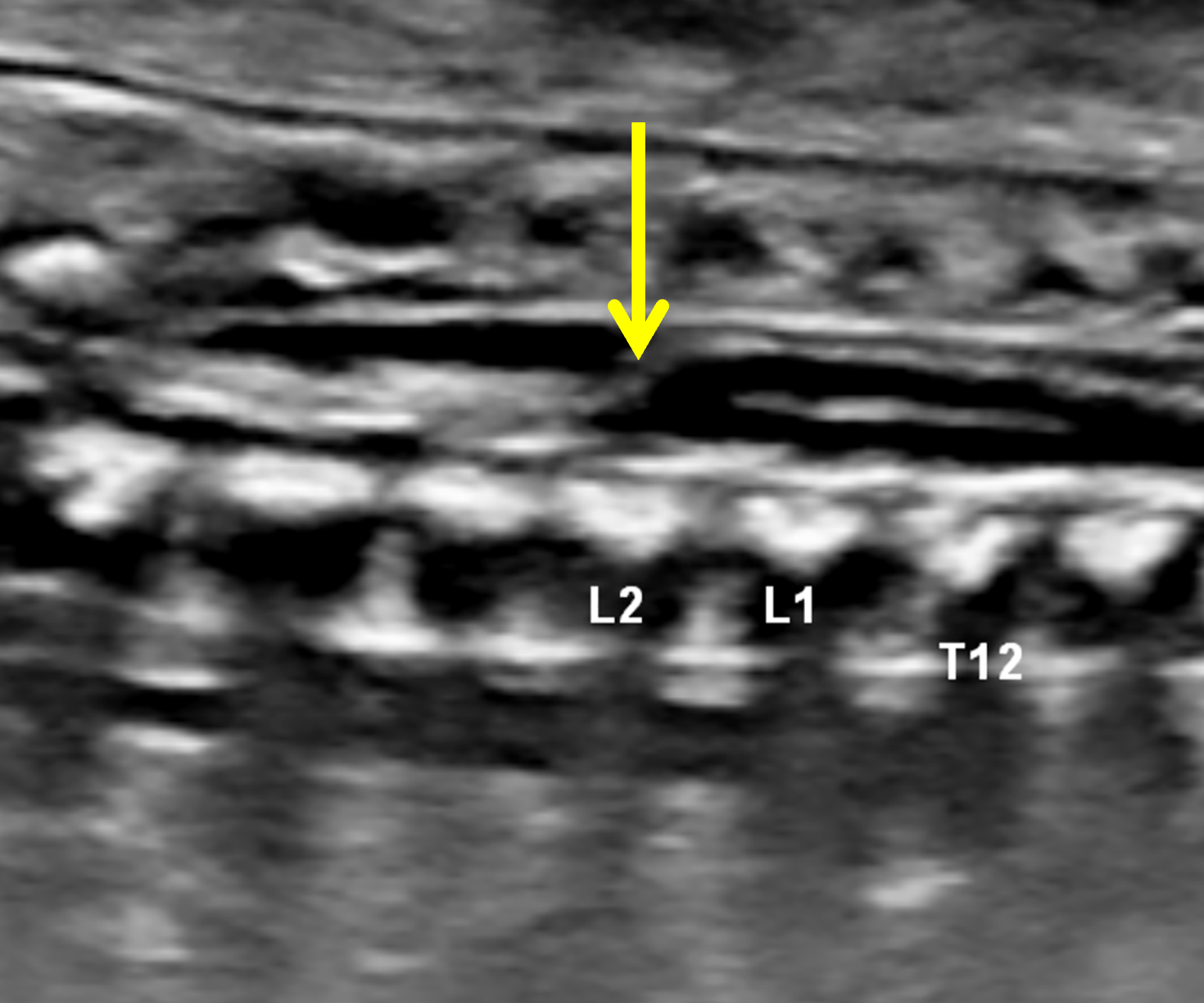
Figure 4: Prenatal diagnosis of caudal regression syndrome at 22 weeks + 6 days gestational age. (4A) Ultrasound image in the sagittal plane of the lower fetal spine shows agenesis of the coccyx and partial agenesis of the sacrum, which ends at S2 (arrow). (4B) Sagittal ultrasound image through the fetal thoracolumbar junction shows the conus medullaris terminating at L2, cranial to its expected position at this stage of development, with a blunted configuration (arrow). Images courtesy of Edward Oliver, MD, PhD, Children’s Hospital of Philadelphia.
Clinical Presentation
The clinical presentation of CRS is variable and depends on the severity of spinal, neural and associated visceral abnormalities.4 Patients with mild forms may remain asymptomatic or present later in childhood with gait abnormalities, genitourinary dysfunction, or constipation. More advanced cases are frequently diagnosed prenatally or shortly after birth because of associated lower extremities deformities or neurologic deficits.5 The most severe forms that extend into the thoracic spine are commonly associated with cardiac or respiratory problems that can lead to early neonatal death.6
On physical examination, patients with sacral agenesis often have a narrow pelvis and hips and flattened buttocks due to gluteal muscle hypoplasia. Lower extremity muscle wasting is also common.4 Sensorimotor deficits are frequently present, with motor impairment typically more pronounced than sensory deficits.7
Epidemiology and Etiology
CRS is a rare disorder with an estimated incidence of 1-2 per 100,000 live births, although the true incidence is likely underestimated as mild cases may go undiagnosed.8 Most cases are sporadic, though familial cases have been reported. There appears to be overlap in genetic pathways implicated in CRS and other developmental disorders, including the VACTERL (vertebral defects, anal atresia, cardiac defects, trachea-esophageal fistula, renal anomalies and limb abnormalities) association.9 There is no sex predilection.
The strongest known risk factor is pregestational maternal diabetes mellitus, which substantially increases the risk of CRS.10 Approximately 15-25% of mothers of children with CRS have insulin dependent diabetes mellitus (Figure 5).11 Conversely, the risk of bearing a child with CRS is reported as high as 1% in diabetic mothers, especially those with poor glycemic control.12
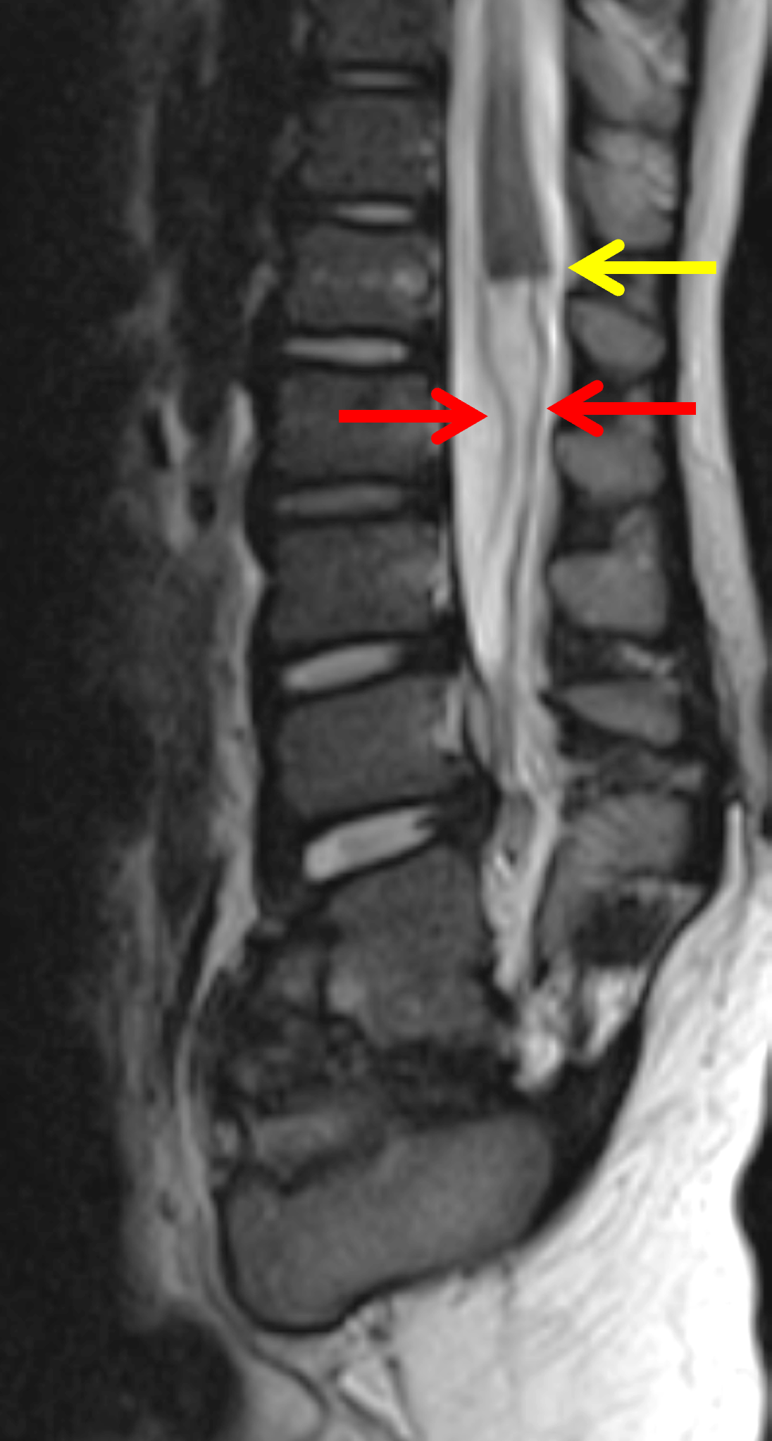
Figure 5: Caudal regression syndrome in a young child with urinary incontinence and multiple congenital anomalies. Maternal history included diabetes mellitus, the strongest recognized prenatal risk factor for CRS. Sagittal T2-weighted image demonstrates partial sacral agenesis, and a truncated, blunted conus medullaris (yellow arrow) with a double-bundle configuration of the cauda equina nerve roots (red arrows). Image courtesy of Franz J. Wippold, M.D.
Imaging Findings
Imaging evaluation of CRS focuses on determining the extent of vertebral regression, assessing spinal cord morphology, identifying associated spinal abnormalities, and evaluating related visceral abnormalities.
Radiography
Radiographs demonstrate the characteristic skeletal abnormalities associated with CRS. The defining finding is partial or complete absence of the sacrum (Figure 2C), with severity ranging from isolated coccygeal agenesis to more extensive lumbosacral regression. Additional findings may include vertebral segmentation anomalies, spinal dysraphism, and scoliosis. The Renshaw Classification categorizes sacral agenesis into four patterns based on the extent of sacral deficiency and the relationship between the remaining spine and pelvis.13 While this classification is useful for describing skeletal abnormalities, it does not reliably predict neurologic function.
CT
CT has a supplemental role in the evaluation of CRS, primarily serving as a tool for characterization of complex skeletal abnormalities rather than initial diagnosis. High-resolution CT with multiplanar and three-dimensional reconstructions allow detailed delineation of vertebral anomalies, pelvic morphology, and the alignment of the spine with the pelvis, particularly in patients undergoing preoperative assessment.
MRI
MRI plays a central role in the evaluation of CRS by providing detailed characterization of neural abnormalities, which are important determinants of neurologic function and long-term impairment. Pang et al. divided patients into two broad groups based on the location and morphology of the conus medullaris.7 This classification may aid in prognostication and surgical planning.
In Group 1, the spinal cord terminates above the L1 level with a shortened, blunted, or truncated appearance (Figure 6). On sagittal images, the distal end of the cord may be wedge-shaped, with the dorsal margin extending more inferiorly than the ventral portion (Figures 2A and 3A).1,3 Additionally, the terminal dorsal and ventral nerve roots are characteristically separated from each other into two distinct bundles (Figures 2-4).1,14 Group 1 patients typically have more extensive sacral deformities, and neurologic deficits present at birth are generally nonprogressive.
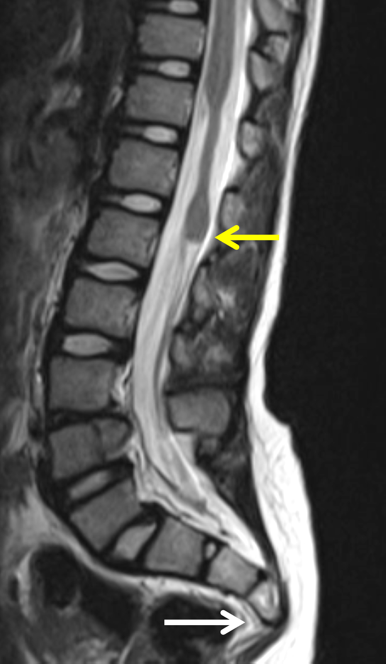
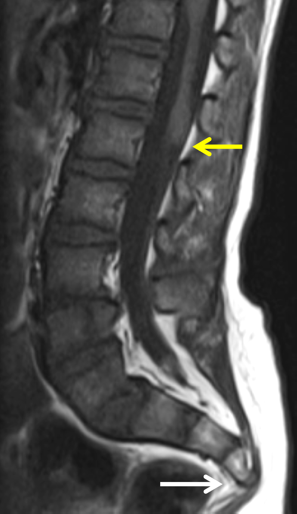
Figure 6: Pang Classification Group 1 caudal regression in a 7-year-old with neurogenic bladder and gait-disturbances. Sagittal (6A) T2-weighted and (6B) T1-weighted images reveal partial sacral agenesis (white arrows) with abnormal morphology of the distal spinal cord, which ends abruptly with a blunted appearance (yellow arrows).
Group 2 patients demonstrate an elongated, thickened, low-lying conus medullaris consistent with tethered cord (Figure 7), and may develop progressive neurologic impairment related to cord tethering. Surgical untethering may be considered in symptomatic patients or those with evidence of neurologic deterioration.7,14
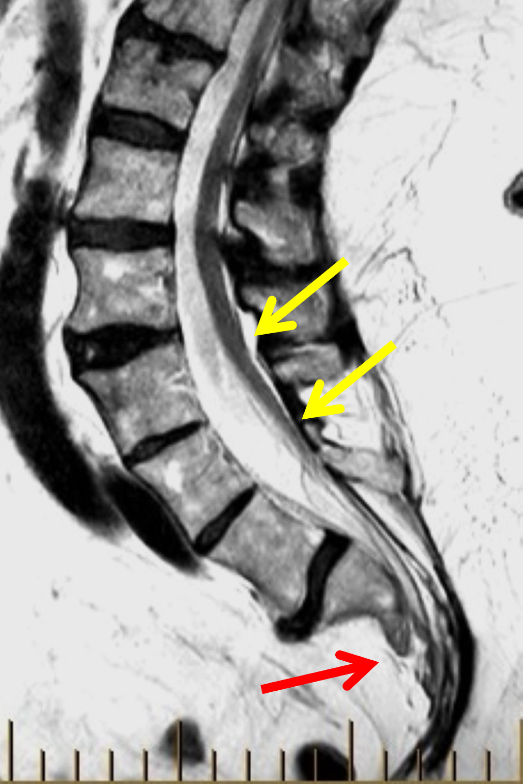
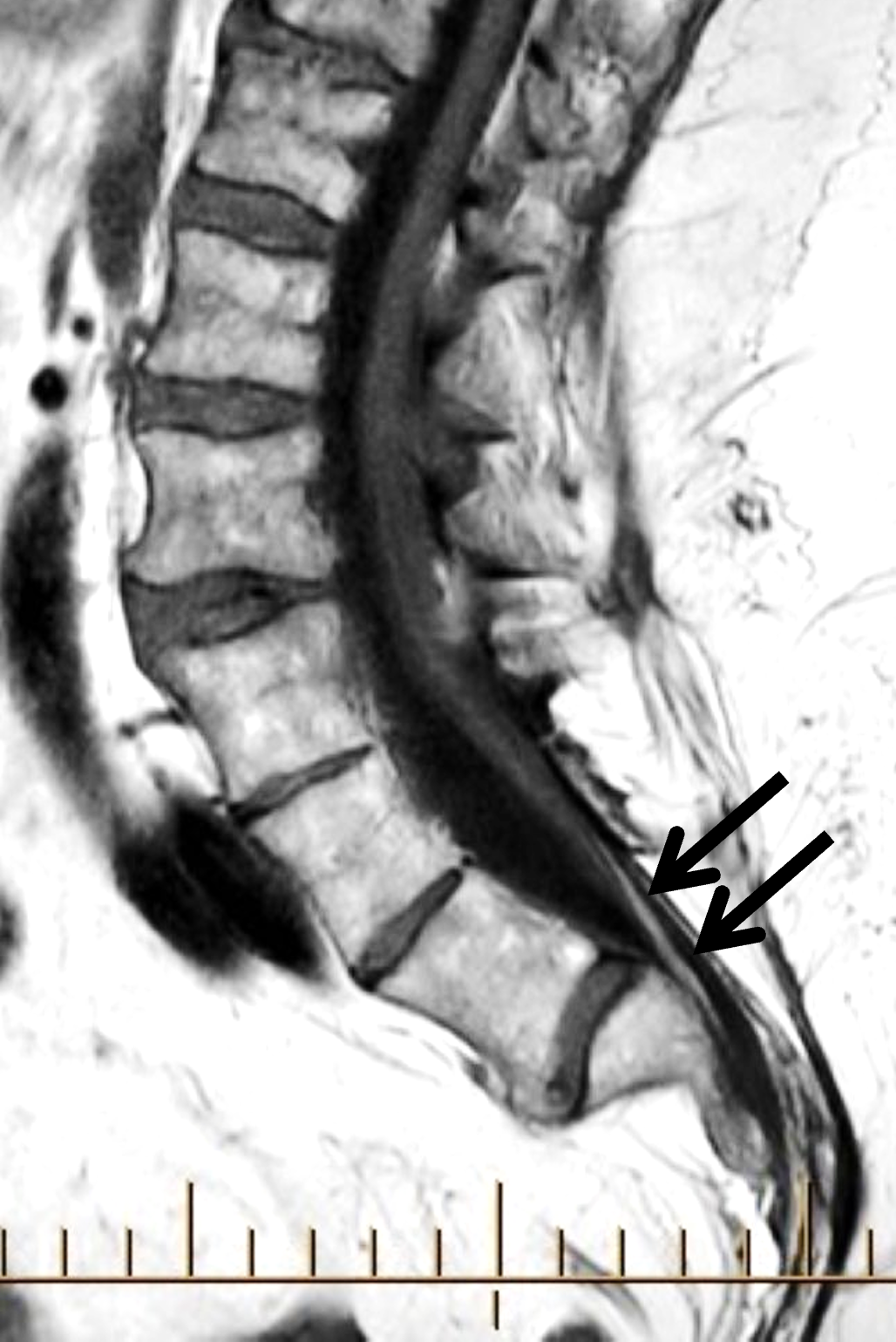

Figure 7: Pang Classification Group 2 caudal regression incidentally found in a 79-year-old man undergoing evaluation for low back pain. (7A) Sagittal T2-weighted image shows partial sacral agenesis (red arrow) and a low-lying conus medullaris with tethering of the spinal cord (yellow arrows). (7B) Sagittal T1-weighted image demonstrates an associated lipoma of the filum terminale (arrows). (7C) Axial T1-weighted imaging confirms partial agenesis of the sacrum (S).
Treatment and Prognosis
Management of CRS requires a multidisciplinary approach tailored to the spectrum of congenital abnormalities and guided by the extent of caudal regression and associated complications.15 Neurologic management focuses on the evaluation and treatment of spinal cord abnormalities, including tethered cord, spinal dysraphism, and scoliosis, with surgical intervention considered in patients with progressive neurologic deterioration. Urologic surveillance and management are critical, as neurogenic bladder dysfunction may lead to progressive renal impairment. Treatment strategies include intermittent catheterization, serial urodynamic assessment, and pharmacologic bladder therapy to preserve renal function and optimize urinary outcomes.16 Gastrointestinal management is directed toward any specific anorectal anomaly present, as well as treatment of constipation or incontinence, and includes surgical and non-surgical interventions.17 Orthopedic management addresses the associated musculoskeletal abnormalities, including hip instability, joint contractures, and foot deformities, with treatment ranging from physical therapy and bracing to complex reconstructive procedures.18
Differential Diagnosis
The differential diagnosis of CRS includes disorders of the caudal spine, pelvis and lower extremities. Accurate differentiation is important because associated abnormalities, prognosis and management differ among these entities.
Currarino Syndrome
Currarino syndrome is a distinct congenital disorder that shares a common embryologic original with CRS. Although both disorders involve abnormal development of the caudal mesoderm and may present with sacral anomalies and spinal dysraphism, Currarino syndrome is characterized by the triad of partial sacral agenesis (typically a hemisacrum), anorectal malformation, and a presacral mass (Figure 8).19 The presacral masses in Currarino syndrome are most commonly anterior sacral meningoceles, teratomas, or enteric cysts. Less common presacral lesions include lipomas, dermoid and epidermoid cysts. Currarino syndrome is an autosomal dominant inherited syndrome caused by defects of the MNX1 homeobox gene (earlier termed HLXB9) on chromosome 7q36.20

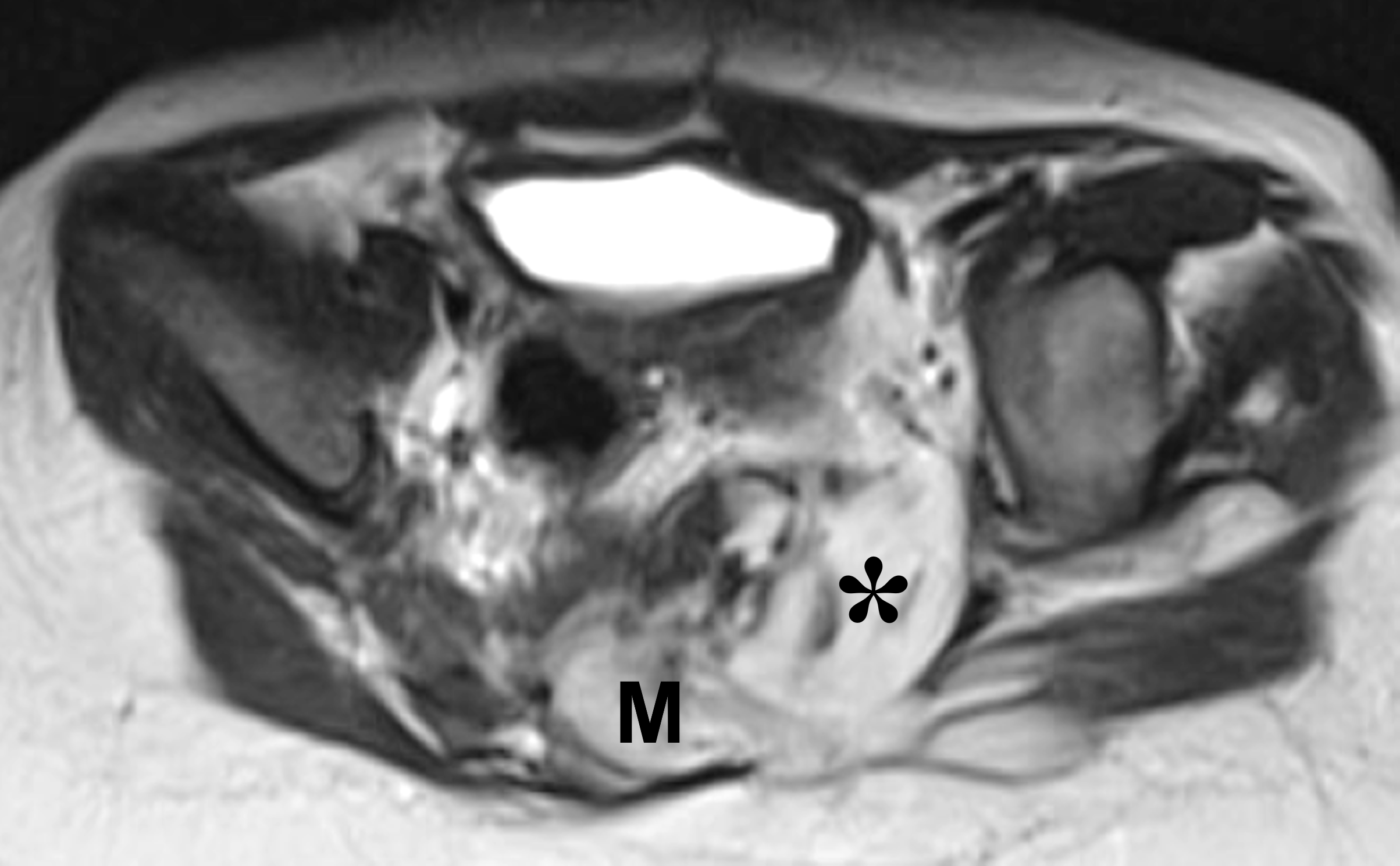
Figure 8: Currarino syndrome in an 8-month-old boy with anal atresia. (8A) Sagittal T2-weighted image shows partial sacral agenesis (S). The cord appears tethered and the distal nerve roots are contained in a lipomeningocele (M). A presacral mass (asterisk) composed predominantly of macroscopic fat is contiguous with the lipomeningocele. (8B) Axial T2-weighted image shows the lipomeningocele (M) and adjacent fatty mass (asterisk).
Sirenomelia
Sirenomelia (mermaid syndrome) is a rare congenital anomaly that overlaps with CRS in its caudal developmental abnormalities and association with maternal diabetes. Sirenomelia, however, is distinguished from typical CRS by fusion of the lower extremities and severe visceral anomalies, most notably bilateral renal agenesis or severe renal dysgenesis.21
VACTERL Association
VACTERL association should be considered in the differential diagnosis because of its overlapping vertebral, anorectal, renal, and limb anomalies. Unlike CRS, however, VACTERL association encompasses a wider spectrum of congenital malformations, particularly cardiac defects and tracheoesophageal anomalies, and does not typically demonstrate the characteristic sacral agenesis and caudal spinal dysgenesis that define CRS.22
Conclusion
Caudal regression syndrome (CRS) is a rare congenital disorder characterized by variable degrees of caudal spinal dysgenesis and a broad spectrum of associated neurologic, genitourinary, gastrointestinal, and musculoskeletal abnormalities. The extent of neural involvement is a key determinant of neurologic function and long-term clinical outcome. Recognition of the characteristic imaging features of CRS and its associated anomalies is essential for accurate diagnosis, differentiation from other caudal developmental disorders, appropriate prognostic counseling, and coordinated multidisciplinary management.
References
- Nievelstein RA, Valk J, Smit LM, Vermeij-Keers C. MR of the caudal regression syndrome: embryologic implications. AJNR Am J Neuroradiol. 1994;15(6):1021–1029. ↩
- Qudsieh H, Aborajooh E, Daradkeh A. Caudal regression syndrome: Postnatal radiological diagnosis with literature review of 83 cases. Radiol Case Rep. 2022;17(12):4636–4641. doi:10.1016/j.radcr.2022.09.037 ↩
- Barkovich AJ, Raghavan N, Chuang S, Peck WW. The wedge-shaped cord terminus: a radiographic sign of caudal regression. AJNR Am J Neuroradiol. 1989;10(6):1223–1231. ↩
- Lee JY, Shim Y, Wang KC. Caudal Agenesis : Understanding the Base of the Wide Clinical Spectrum. J Korean Neurosurg Soc. 2021;64(3):380–385. doi:10.3340/jkns.2021.0025 ↩
- Krishnan V, Jaganathan S, Jayappa S, Glasier C, Choudhary A, Ramakrishnaiah R, et al. Clinical and radiological evaluation of caudal regression syndrome. Pediatr Radiol. 2024;54(9):1451–1461. doi:10.1007/s00247-024-05945-1 ↩
- Sen KK, Patel M. Caudal Regression Syndrome. Med J Armed Forces India. 2007;63(2):178–179. doi:10.1016/S0377-1237(07)80071-2 ↩
- Pang D. Sacral agenesis and caudal spinal cord malformations. Neurosurgery. 1993;32(5):755–778; discussion 778–759. doi:10.1227/00006123-199305000-00009 ↩
- Bhatt S, Tandon A, Kumar Singh A, Manchanda S, Jain S, Meena N. Caudal Regression Syndrome:A Case Study With Associated Review of Common Differential Diagnoses Made With Antenatal Sonography. Journal of Diagnostic Medical Sonography. 2017;33(2):130–133. doi:10.1177/8756479316677012 ↩
- Warner T, Scullen TA, Iwanaga J, Loukas M, Bui CJ, Dumont AS, et al. Caudal Regression Syndrome-A Review Focusing on Genetic Associations. World Neurosurg. 2020;138461–467. doi:10.1016/j.wneu.2020.03.057 ↩
- Boulas MM. Recognition of caudal regression syndrome. Adv Neonatal Care. 2009;9(2):61–69; quiz 70–61. doi:10.1097/ANC.0b013e31819de44f ↩
- Al Kaissi A, Klaushofer K, Grill F. Caudal regression syndrome and popliteal webbing in connection with maternal diabetes mellitus: a case report and literature review. Cases J. 2008;1(1):407. doi:10.1186/1757-1626-1-407 ↩
- Palacios-Marqués A, Oliver C, Martín-Bayón T, Martinez-Escoriza JC. Prenatal diagnosis of caudal dysplasia sequence associated with undiagnosed type I diabetes. BMJ Case Rep. 2013;2013. doi:10.1136/bcr-2013-009043 ↩
- Renshaw TS. Sacral agenesis. J Bone Joint Surg Am. 1978;60(3):373–383. ↩
- Puneeth KT, Goyal A, Jana M. High abrupt cord termination: a hallmark of caudal regression syndrome. BMJ Case Rep. 2014;2014. doi:10.1136/bcr-2013-201770 ↩
- Sánchez-Romero M, Tlaxcala-Castillo L, Pichardo-Rojas PS, Valencia-Melo MA, Paz-López Á A, Sánchez-Sagastegui F, et al. Sacral Agenesis. Pediatr Neurol. 2025;16327–34. doi:10.1016/j.pediatrneurol.2024.10.020 ↩
- Esposito G, Totonelli G, Iacobelli BD, Longo D, Caldaro T, Blasetti G, et al. Continence management in children with severe caudal regression syndrome: role of multidisciplinary team and long-term follow-up. Pediatr Surg Int. 2022;38(10):1461–1472. doi:10.1007/s00383-022-05168-1 ↩
- Theeuwes JN, de Beaufort CMC, McDonald CJ, Bakker DP, van Schuppen J, Kuijper CF, et al. Bowel and bladder outcomes in patients with anorectal malformations and sacral agenesis: a retrospective cohort study. Pediatr Surg Int. 2026;42(1). doi:10.1007/s00383-026-06471-x ↩
- Dumont CE, Damsin JP, Forin V, Carlioz H. Lumbosacral agenesis. Three cases of reconstruction using Cotrel-Dubousset or L-rod instrumentation. Spine (Phila Pa 1976). 1993;18(9):1229–1235. ↩
- Currarino G, Coln D, Votteler T. Triad of anorectal, sacral, and presacral anomalies. AJR Am J Roentgenol. 1981;137(2):395–398. doi:10.2214/ajr.137.2.395 ↩
- Dworschak GC, Reutter HM, Ludwig M. Currarino syndrome: a comprehensive genetic review of a rare congenital disorder. Orphanet J Rare Dis. 2021;16(1):167. doi:10.1186/s13023-021-01799-0 ↩
- Stocker JT, Heifetz SA. Sirenomelia. A morphological study of 33 cases and review of the literature. Perspect Pediatr Pathol. 1987;107–50. ↩
- Solomon BD. VACTERL/VATER Association. Orphanet J Rare Dis. 2011;656. doi:10.1186/1750-1172-6-56 ↩
