
Clinical History
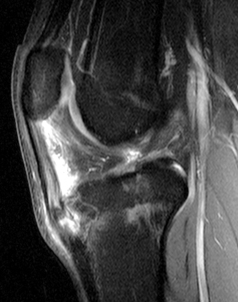
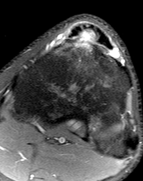
A 16-year-old boy, active in sports, complains of anterior knee pain and swelling located inferior to the patella, which increases with activity. Sagittal (1A) T1-weighted and (1B) fat-suppressed, fluid-sensitive images, (1C) a lateral radiograph, and (1D) an axial fat-suppressed, fluid-sensitive image are shown. What are the findings? What is your diagnosis?


Findings

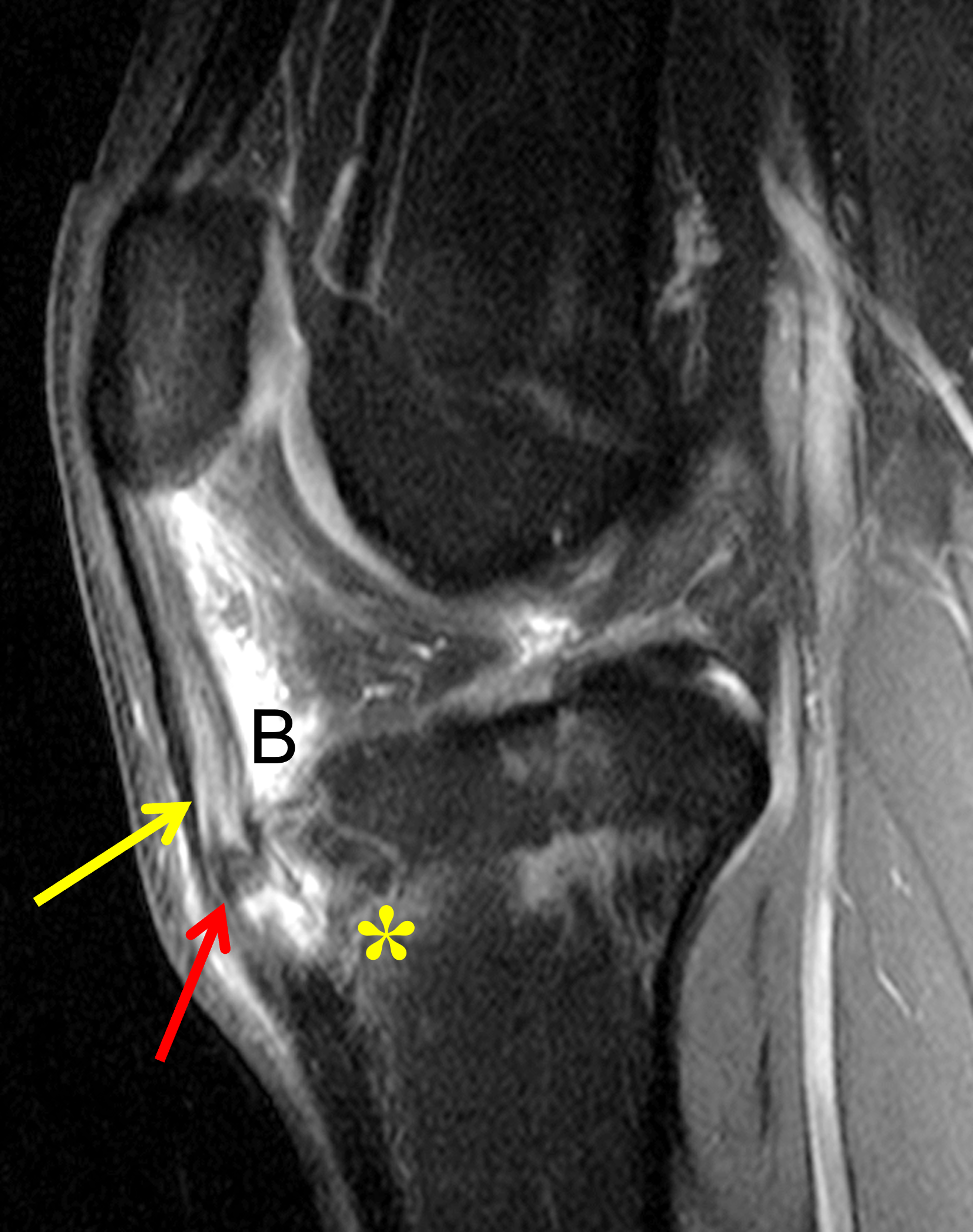

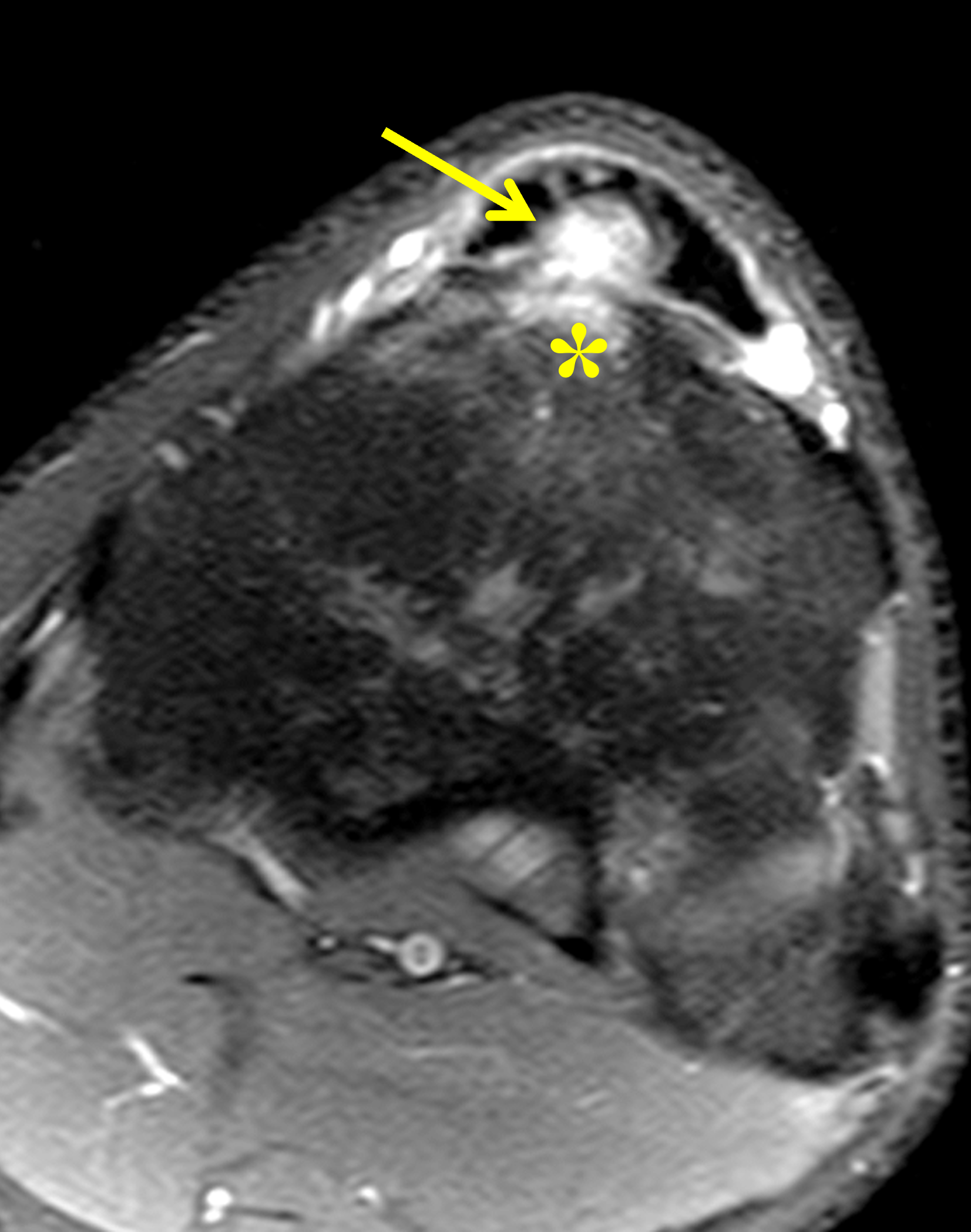
Figure 2: (2A) Sagittal T1-weighted image shows thickening and increased signal in the distal patellar tendon (yellow arrow) containing a low signal-intensity ossicle (red arrow), with associated hypertrophy of the anterior tibial tubercle (black arrow). (2B) Sagittal fat-suppressed, fluid-sensitive image shows increased signal in the distal patellar tendon (yellow arrow) with slight marrow edema in the ossicle (red arrow), and marrow edema in the underlying tibia (asterisk). Fluid distends the deep infrapatellar bursa (B). (2C) Lateral radiograph demonstrates patella alta with a thick, indistinct patellar tendon (yellow arrow) containing a mature ossification distally (red arrow), as well as hypertrophic bone changes affecting the tibial tuberosity (black arrow). (2D) Axial fat-suppressed, fluid-sensitive image redemonstrates the marrow edema in the anterior tibia (asterisk) and the high signal within the distal patellar tendon (arrow).
Diagnosis
Osgood-Schlatter disease, active phase.
Introduction
Osgood-Schlatter disease, first described independently by Osgood in Boston and Schlatter in Zurich in 1903, is an overuse traction apophysitis of the anterior tibial tuberosity (tubercle). It affects approximately 1 in 10 preadolescent and adolescent athletes and typically presents with anterior knee pain and swelling.1,2
Etiology
Osgood and Zurich believed that Osgood-Schlatter disease resulted from avulsion of a portion of the anterior tibial tuberosity apophysis due to indirect trauma caused by quadriceps contraction. Some later authors proposed osteonecrosis as the primary etiology of Osgood-Schlatter disease, though subsequent studies have shown that the anterior tibial tuberosity has an excellent blood supply. Findings from a small-sample pathologic study were most supportive of a traumatic etiology, with injury occurring somewhere along the attachment of the patellar tendon to the anterior tibial tuberosity.1-3
While some investigators have reported that injury to the anterior tibial tuberosity ossification center is the primary process, with associated secondary inflammatory changes of the patellar tendon and adjacent soft tissues, others have proposed that overuse-related inflammatory changes in the soft tissues, specifically the insertional patellar tendon, are the primary process, with secondary adjacent osseous changes. Though these theories have been debated, it seems likely that the main etiologic factor is repetitive tensile stress on the distal patellar tendon and tendon enthesis, resulting in a micro-traumatic insult to the distal tendon and/or tendinous insertion site, potentially associated with small chondral/osteochondral avulsion fractures and/or metaplastic intra-tendinous ossicle formation.4,5
Over the years, many potential risk factors for, or associations with, the development of Osgood-Schlatter disease have been studied, including variations of patellofemoral alignment and anatomy, decreased elasticity of the patellar tendon, relative quadriceps and hamstring muscle strength and tightness, increased posterior tibial slope, elevated tibial torsion angles, limited ankle dorsiflexion, body weight, early sport specialization and, interestingly, a diagnosis of attention-deficit hyperactivity disorder (ADHD).6,7,8,9 Quadriceps muscle tightness, reduced hamstring flexibility, greater body weight, and higher BMI were identified as risk factors by Nakase et al. in a 2015 prospective cohort study of 300 knees.7 Although the precise contribution of these reported risk factors remains controversial and incompletely understood, current prevention strategies emphasize balanced training and cross-training, as well as regular warm-up regimens that incorporate quadriceps and hamstring stretching.6,7,10
Clinical Presentation and Diagnosis
Osgood-Schlatter disease is estimated to affect about 1 out of 10 adolescents. It typically occurs between ages 11 and 15 in boys, with an earlier onset in girls. Osgood-Schlatter disease was initially reported to be more common in males. However, current studies show no male predilection, possibly due to greater female participation in high-impact sports in recent years. The condition is bilateral in up to 25% of cases. Most affected individuals participate in athletic activities that involve jumping, kicking, running, or squatting. Symptoms often develop during or immediately after a period of rapid growth.1,6,11
Patients typically complain of anterior knee pain and swelling, often aggravated by activity, knee extension, and kneeling, and improved with rest. Symptoms may initially be intermittent and mild, becoming more severe and continuous over time. Clinical examination reveals soft tissue swelling, sometimes firm or mass-like, in the region of the anterior tibial tuberosity or distal patellar tendon, usually without a joint effusion. Resisted knee extension may reproduce or aggravate the anterior pain.
Imaging Findings
Though the diagnosis of Osgood-Schlatter disease may be made based on clinical findings alone, screening radiographs are often obtained in an athlete with ongoing pain, primarily to exclude alternative pathology. In patients with Osgood-Schlatter disease, the lateral projection is most informative, and it is important that the lateral radiograph not be over-penetrated, as this may obscure subtle soft-tissue findings. A lateral view obtained with slight internal rotation will optimally depict the anterior tibial tuberosity.1
With active Osgood-Schlatter disease, lateral knee radiographs usually show soft-tissue swelling anterior to the anterior tibial tuberosity. Increased radiodensity in the distribution of Hoffa’s fat pad, thickening of the distal patellar tendon contour, and an indistinct posterior margin of the distal patellar tendon may be seen (Figure 3). In young patients when the anterior tibial tuberosity apophysis is entirely cartilaginous, no bone findings will be evident. In older patients with active disease, in addition to the soft tissue findings, one or more ossicles anterior to the anterior tibial tuberosity may be present. These bone fragments may represent dystrophic heterotopic ossification in the inflamed patellar tendon, or fragmentation of the apophysis. With time, the ossicles may coalesce with the underlying bone, contributing to anterior tibial tuberosity hypertrophy.1
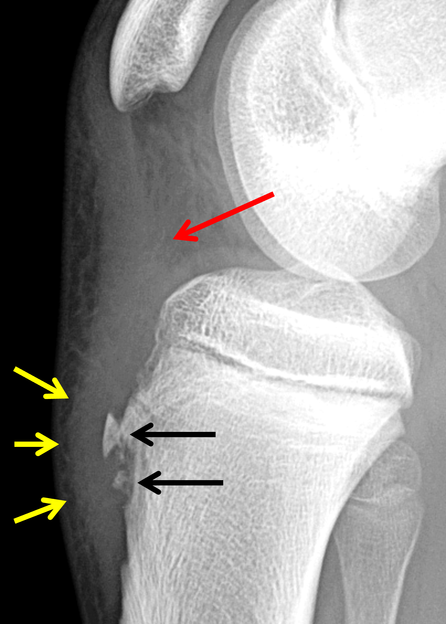
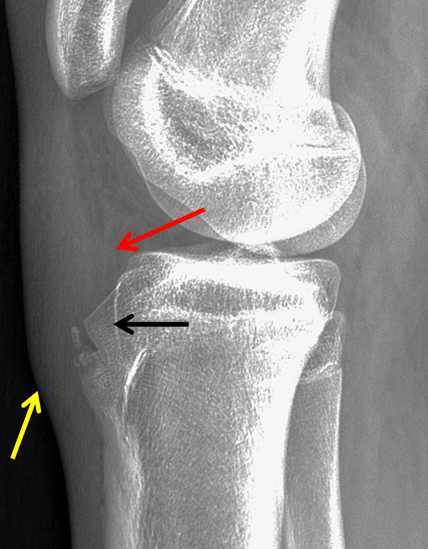
Figure 3. Active Osgood-Schlatter disease in two patients with anterior knee pain. Lateral radiographs. (3A) 12-year-old boy showing soft tissue swelling anterior to the tibial tubercle (yellow arrows) and an indistinct posterior margin of the distal patellar tendon where it abuts Hoffa’s fat pad (red arrow). Small ossicles representing fragmentation of the anterior tibial tubercle apophysis and heterotopic ossification in the patellar tendon are visible (black arrows). (3B) 13-year-old girl shows a later stage of skeletal maturity compared to Figure 3A. Note the soft tissue swelling (yellow arrow) anterior to the fused tibial tubercle apophysis, which shows mild residual fragmentation as well as osseous hypertrophy (black arrow). Soft tissue edema in Hoffa’s fat pad contributes to the indistinct contour of the distal patellar tendon (red arrow).
It is important to recognize that one or more ossification centers of the anterior tibial tuberosity may normally be present in girls between the ages of 8 and 12 years and boys between the ages of 9 and 14 years. Fusion of these ossification centers with the anterior tibial metaphysis does not usually occur before the age of 15 years in girls and 17 years in boys. Thus, the presence of anterior ossicles alone in adolescents may be a normal finding and, in the absence of clinical findings or soft tissue abnormalities, should not be deemed pathologic (Figure 4).
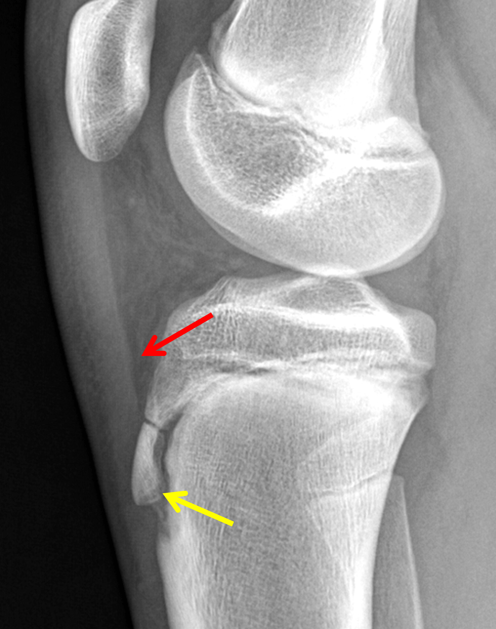
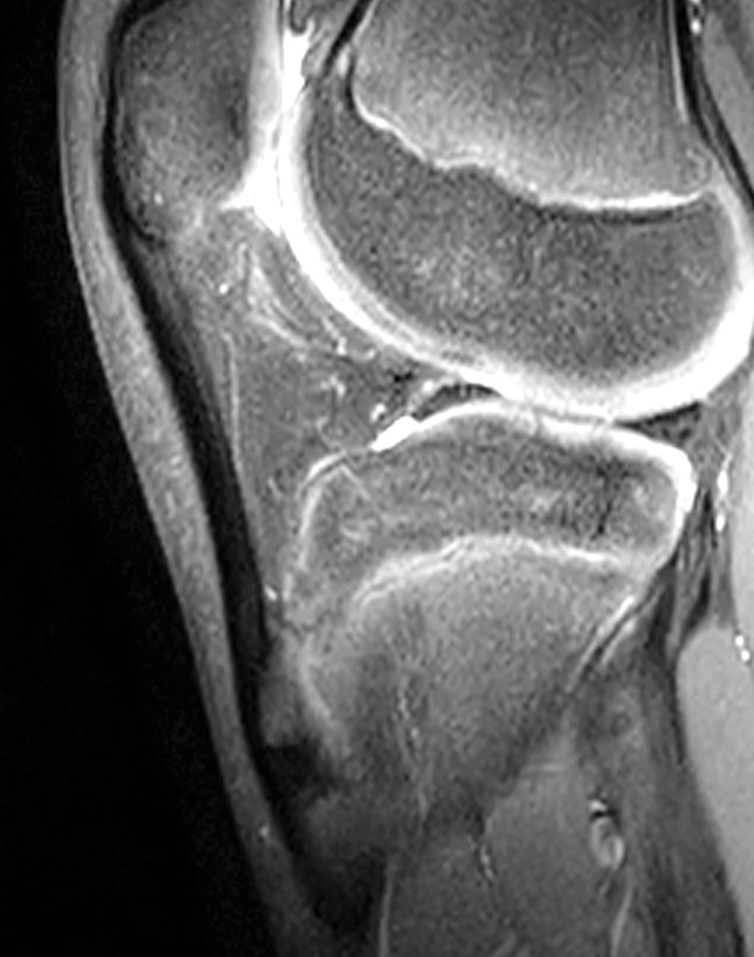
Figure 4. Normal fragmentation of the anterior tibial tuberosity apophysis in a 13-year-old boy with no anterior knee pain. (4A) Lateral radiograph shows a fragmented ossification center in the tibial tuberosity apophysis (yellow arrow). Note the lack of overlying soft tissue swelling and the normal thickness patellar tendon with a sharp posterior margin (red arrow). (4B) Corresponding fat-suppressed, fluid-sensitive sequence shows no marrow edema in the tibial tuberosity or underlying bone, and no soft tissue signal changes in the overlying subcutaneous tissues, patellar tendon, or Hoffa’s fat pad. Contrast with Figure 2B.
MRI, because of its ability to demonstrate abnormalities of both bone and soft tissue structures, is a useful second imaging examination when symptoms are atypical or persistent. Although Doppler ultrasound has been reported to accurately detect active Osgood-Schlatter disease, MRI provides a more comprehensive evaluation. With active Osgood-Schlatter disease, fat-suppressed PD-weighted or T2-weighted images often show marrow edema within the anterior tibial tuberosity (Figure 5) in addition to overlying anterior subcutaneous edema. Soft tissue edema and/or deep infrapatellar bursitis may also occur within Hoffa’s fat pad adjacent to the distal patellar tendon (Figure 6). Thickening of the distal patellar, with heterogeneous increased internal signal on fluid-sensitive sequences and an indistinct posterior margin, is frequently present, representing inflammation (Figure 2). Foci of heterotopic ossification may develop within the diseased patellar tendon and later enlarge. Marrow edema in these ossicles is an additional finding of active disease (Figure 7). Pathologic changes in the anterior tibial tuberosity secondary ossification center can include marrow edema, fragmentation, or actual avulsion of the apophysis; with progressive disease an avulsed fragment can separate and displace slightly superiorly. Early findings in the apophysis may be radiographically occult and visible only on MRI. Over time, the gap between an avulsed chondral or osteochondral fragment and the underlying bone can fill with fibrocartilage, which may or may not subsequently ossify and heal (Figure 8).1,4,5,12

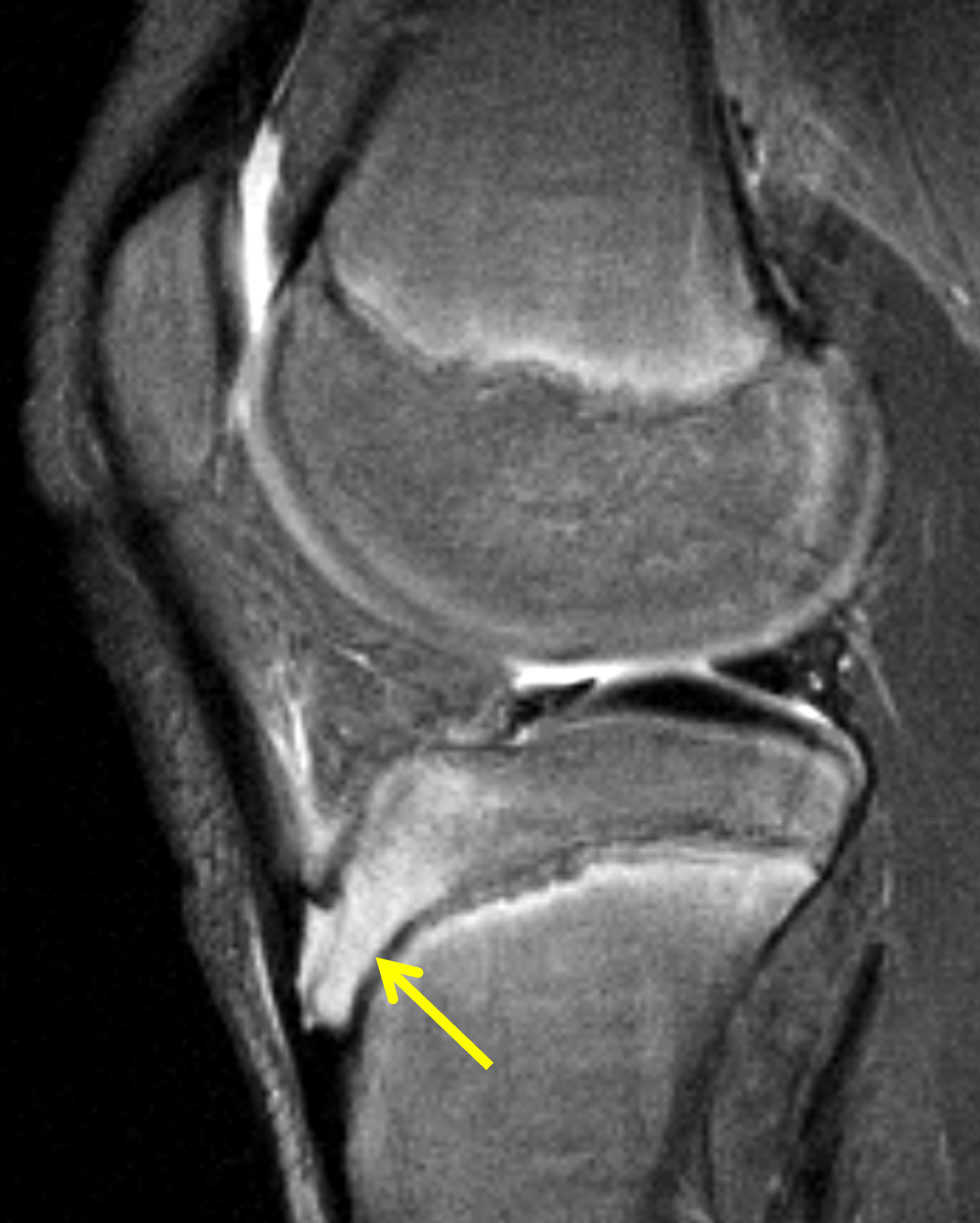
Figure 5. Active Osgood-Schlatter disease in a 12-year-old boy. Sagittal (5A) T1-weighted and (5B) fat-suppressed, fluid-sensitive images demonstrate marrow edema in the anterior tibial tuberosity secondary growth center (arrows).
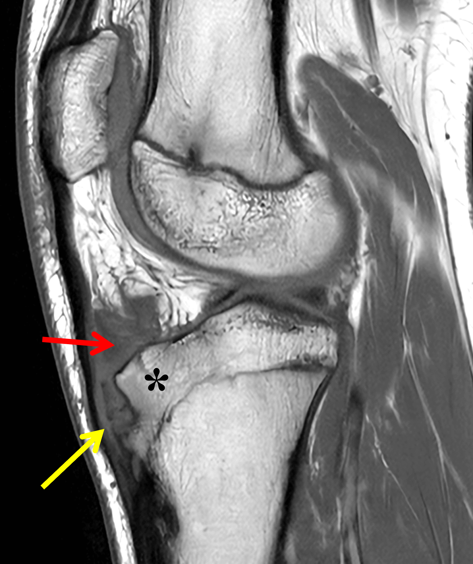
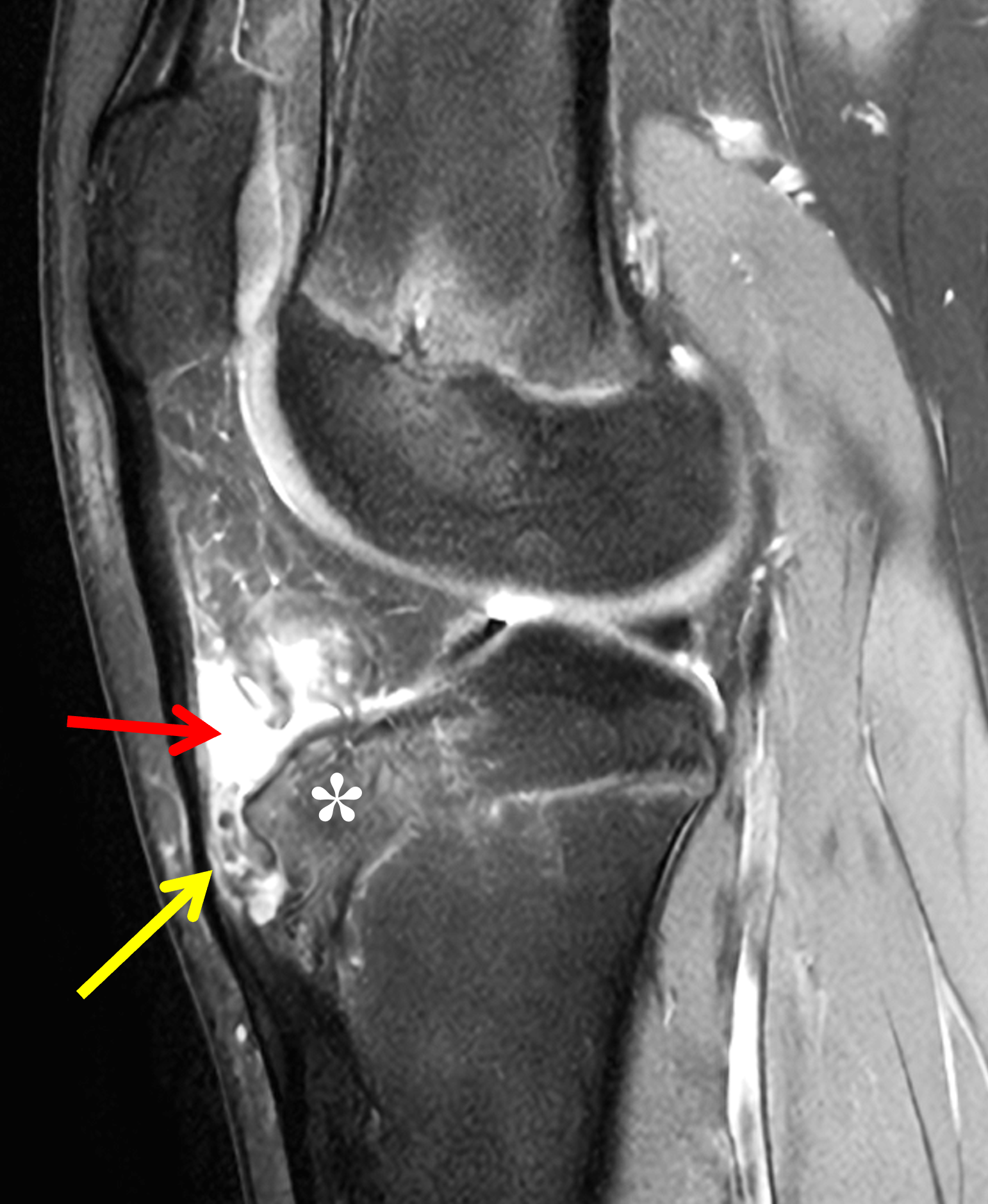
Figure 6. Active Osgood-Schlatter disease in a 13-year-old girl. Sagittal (6A) T1-weighted and (6B) fat-suppressed, fluid-sensitive images show a distended deep infrapatellar bursa in the inferior aspect of Hoffa’s fat pad (red arrows) in addition to marrow edema in the anterior tibial tuberosity (asterisks). Note the multiple small bone fragments anterior to the tubercle (yellow arrows).
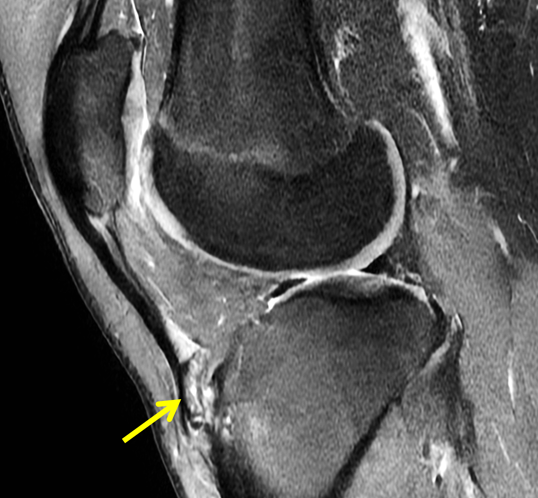
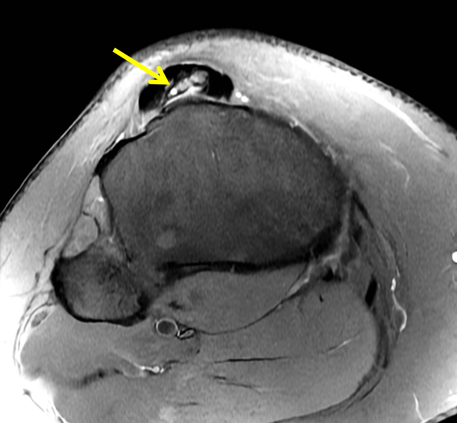
Figure 7. Osgood-Schlatter disease in a 17-year-old boy, with residual/recurrent symptoms. (7A) Sagittal and (7B) axial fat-suppressed, fluid-sensitive images show a large ossification in the distal patellar tendon (arrows). Marrow edema within the ossification is often associated with active disease and symptoms.

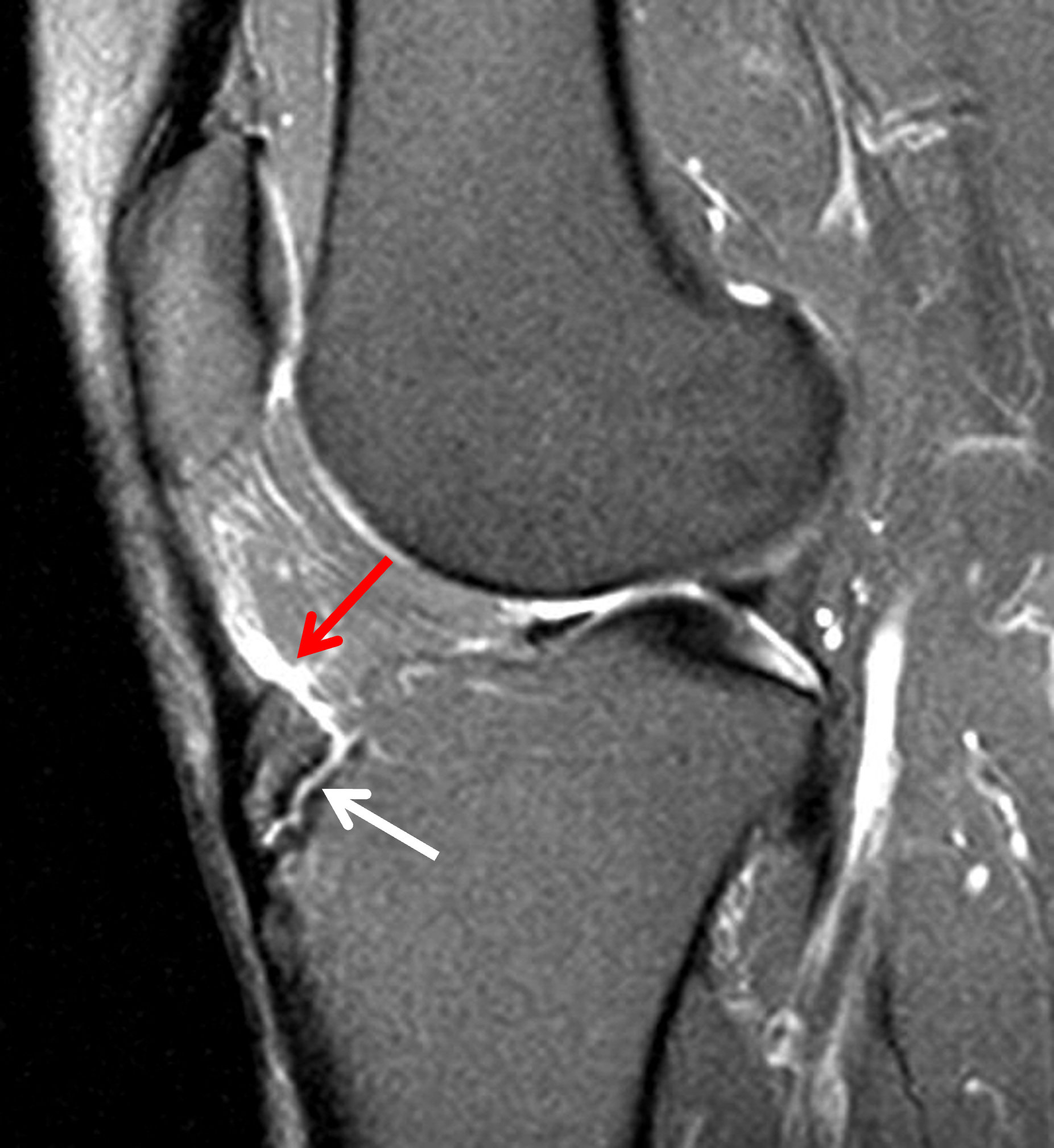
Figure 8. Sequela of childhood Osgood-Schlatter disease in an adult. Inactive disease. (8A) Sagittal T1-weighted image shows an united, avulsed fragment of the anterior tibial tuberosity (arrow). (8B) Sagittal fat-suppressed, T2-weighted image shows a high-signal interface between the bone fragment and underlying tibia (white arrow). Despite the soft edema in the inferior aspect of Hoffa’s fat pad (red arrow) and the minimal marrow edema in the avulsed fragment and tibia tubercle, the patient had no anterior knee pain or symptoms.
A study by Hirano et al.5 evaluated 40 knees with clinically diagnosed Osgood-Schlatter disease and longitudinally followed 22 patients with serial MRI examinations. They classified the MRI features of Osgood-Schlatter disease into five stages (normal, early, progressive, terminal, and healing) and correlated imaging findings with symptomatology. In clinical practice, this degree of detailed staging is not as important as identifying findings indicating active disease (edematous bone and soft tissue structures) versus inactive disease. However, diagnosis of active disease also requires positive clinical findings because radiographic and MRI findings typically lag clinical improvement. Furthermore, imaging abnormalities may only partially resolve despite complete symptom resolution, and healing at the interface between the ossicle(s) and the anterior tibial tubercle is not necessary for symptom abatement.4,5
In adults, it is not uncommon to observe ossification in the distal patellar tendon and/or overgrowth of the anterior tibial tuberosity with no associated symptoms, representing sequela of childhood Osgood-Schlatter disease (Figure 9). Occasionally, an adult with previously resolved childhood Osgood-Schlatter disease may develop new symptoms due to disease “reactivation.” MRI in these patients may demonstrate signal abnormalities in the distal patellar tendon, marrow edema in tendon ossicles and/or the anterior tibial tubercle, and deep infrapatellar bursitis (Figure 10).
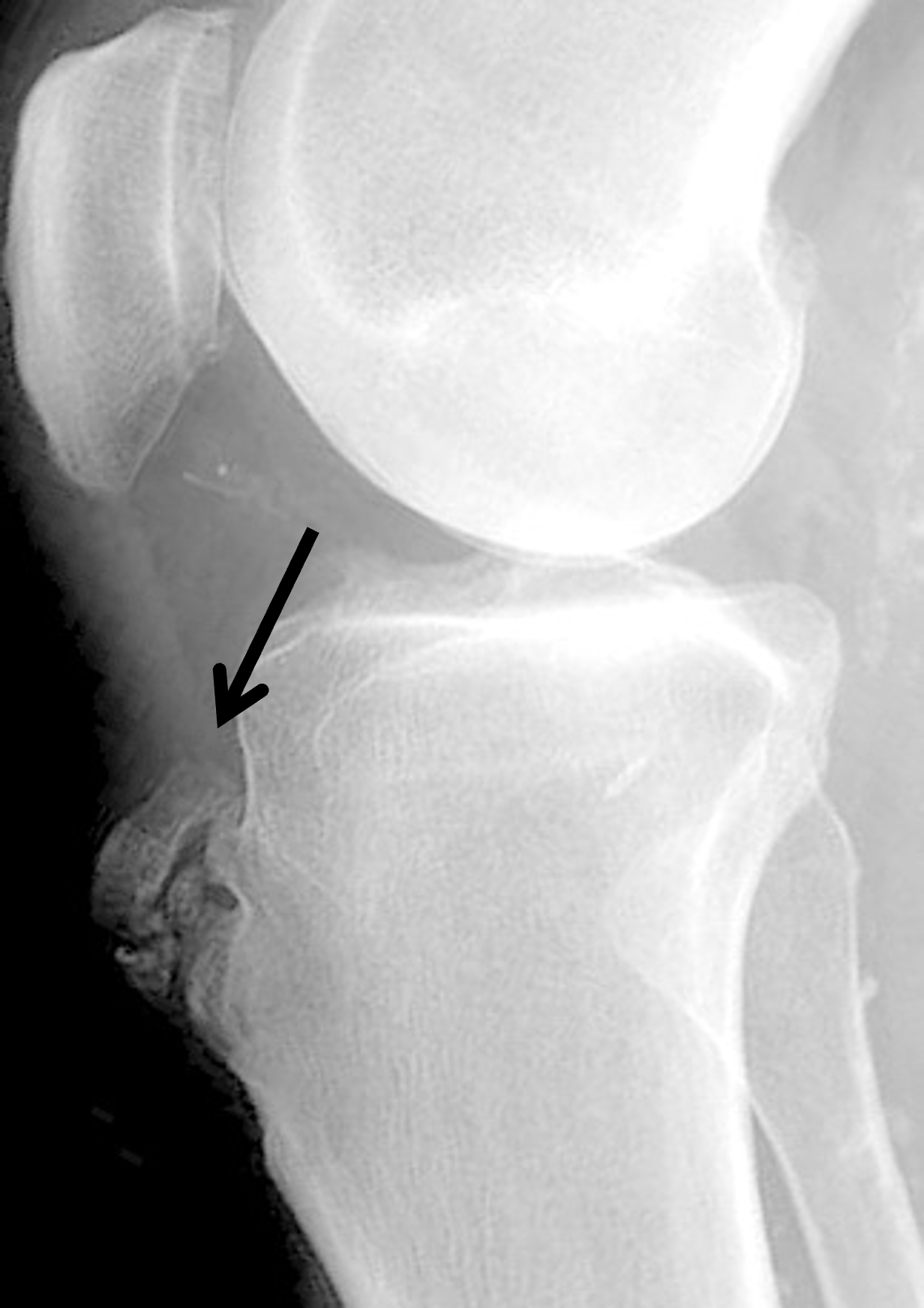


Figure 9. Inactive sequela of childhood Osgood-Schlatter disease in an adult with no associated symptoms. (9A) Lateral radiograph shows mature soft tissue ossification anterior to the tibial tuberosity (arrow). Sagittal (9B) proton density-weighted and (9C) T2-weighted images demonstrate a large, non-edematous ossicle in the distal patellar tendon (arrows).


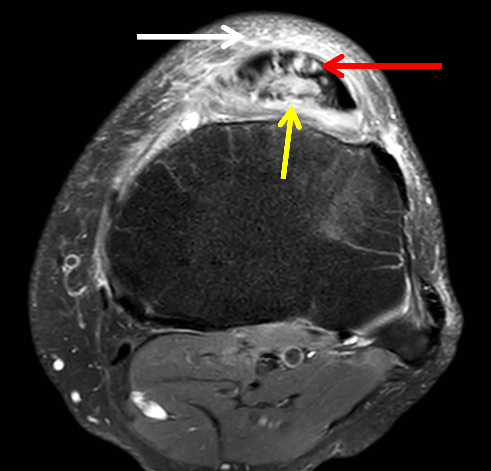

Figure 10. “Reactivation” Osgood-Schlatter disease in a 55-year-old man with severe prepatellar and infrapatellar knee pain. (10A) Sagittal T1-weighted image shows an edematous ossicle (arrow) in the distal patellar tendon. The tendon is thickened, with an indistinct posterior margin (arrowheads). (10B) Sagittal fat-suppressed, fluid-sensitive image shows marrow edema within the ossicle (arrow) and extensive increased signal in the thick patellar tendon (arrowheads) representing active tendinitis. (10C) Axial fat-suppressed, fluid-sensitive image through the proximal tibia shows marrow edema in the distal patellar tendon ossification (yellow arrow), high signal intensity foci in the tendon (red arrow) and severe overlying subcutaneous edema (white arrow). (10D) Axial fat-suppressed, fluid-sensitive image through the patella demonstrates deep partial-thickness and full-thickness degenerative cartilage loss of the medial patellar facet, which may contribute to the patient’s anterior knee pain. Symptoms subsided after surgical excision of the ossicle and soft tissue debridement.
Course, Treatment, and Complications
Historically, Osgood-Schlatter’s Disease was described as an innocuous, mostly self-limited condition, with 80-90% of patients improving with conservative therapy, and gradual symptom resolution upon reaching skeletal maturity.6,11,13,14,15 But recent studies have challenged this assumption. A 2019 retrospective study of 43 patients diagnosed with Osgood-Schlatter disease found that 60% of patients reported persistent knee pain 4 years after diagnosis.9,13 Additionally, even in those who do recover, symptoms may persist for 6 months to 2 years before complete resolution. This prolonged symptomatic period often requires activity modification and multiple time-consuming visits to physicians and physical therapists, which can be challenging for the adolescent and their families, interfering with athletic ambitions and quality of life.8,9,16
Initial conservative management is often guided by clinician experience rather than by evidence-based guidelines. Typical measures include ice, NSAIDs, rest, and activity modification, frequently with a temporary reduction or pause in sports participation. Infrapatellar strapping and tubercle padding may be advised, and a short period of controlled immobilization is sometimes implemented for severe symptoms. Physical therapy focuses on quadriceps and hamstring stretching, progressive knee and hip strengthening, and sometimes core stabilization exercises. Specific rehabilitation protocols and reported success rates vary widely, suggesting the need for future studies to develop and validate evidence-based rehabilitation and strengthening guidelines.6,9,11
Injection-based therapies of the anterior tibial tuberosity, including hyperosmolar dextrose and leukocyte-rich platelet-rich plasma (LR-PRP) injections, have recently been investigated with no reported complications and encouraging results. Although additional randomized controlled studies are needed, preliminary investigations by Guszczyn et al. showed LR-PRP injections to the anterior tibial tuberosity to be an effective and time-efficient alternative to rehabilitation therapy, with improvement in pain and function in reported following a single injection. Injection results are better in patients with a shorter duration of disease.11,16,17
In an estimated 5 to 10% of cases, disabling pain or swelling at the anterior tibial tuberosity persists after skeletal maturity and may interfere with sports, occupational or daily activities, occasionally requiring surgery.15,18 Multiple different surgical interventions for refractory Osgood-Schlatter disease have been described, including open, arthroscopic, and bursoscopic techniques. In 1934, Bosworth et al. described inserting a bone peg into the anterior tibial tubercle to promote ossicle fusion. Although this intervention led to pain resolution, the procedure resulted in a persistent bony prominence.17,19 In 2007, Weiss et al. reported that 80% of patients treated with open ossicle excision, tibial tubercleplasty, and patellar tendon repair were able to return to sports.14,18,20 In 2017, Pagenstert et al. reported improved pain and functional outcomes in seven patients who underwent closing-wedge reduction osteotomy of the prominent anterior tibial tuberosity after failed conservative and prior surgical treatment for recalcitrant Osgood-Schlatter disease.18 Current procedures generally involve excision of ununited ossicles and cartilaginous fragments, patellar tendon debridement and repair, removal of inflamed bursae, and smoothing, resection, or osteotomy of the hypertrophied anterior tibial tuberosity. In most cases, endoscopic surgery is preferred because it is associated with better outcomes and fewer complications than open surgery. However, large ossicles and revision cases may necessitate an open approach.6,11
Osgood-Schlatter disease can be associated with patella alta, possibly related to excessive repetitive tensile forces on the patellar tendon and the anterior tibial tuberosity apophysis.12,21 An underrecognized potential complication of Osgood-Schlatter disease is the development of lateral patellofemoral maltracking, which can lead to accelerated patellofemoral osteoarthritis, with or without patellofemoral instability. In 2021, Kamel et al.12 performed a retrospective study of 171 MRIs in adults with imaging findings of prior Osgood-Schlatter disease, comparing them with a control group of 342 randomly selected MRIs without these findings. The investigators found that 59% of adults with a history of Osgood-Schlatter disease had imaging features suggesting patellofemoral maltracking – which they defined as edema in the superolateral aspect of Hoffa’s fat pad or a TT-TG distance of greater than or equal to 20 mm – compared with 15% in the control group. Patellofemoral arthritis was present in 63% of the Osgood-Schlatter disease cohort.12
The most common long-term complications of Osgood-Schlatter disease are persistent anterior pain and swelling, patellofemoral maltracking, and patellofemoral arthritis. Less commonly reported complications include decreased range of motion and quadriceps wasting related to periods of rest and immobilization, increased tibial slope with potentially associated altered biomechanics, and genu recurvatum.1,11,22
Differential Diagnosis
The main differential diagnoses for anterior, infrapatellar pain and swelling in an adolescent include acute avulsion fracture of the tibial tubercle, tendinosis or tear involving the distal patellar tendon, Hoffa’s syndrome, and infrapatellar bursitis. Tibial tubercle avulsion fractures are uncommon, most often occurring in boys 12-17 years old following forceful quadriceps contraction or knee flexion, as may occur during jumping or sprinting. Unlike Osgood-Schlatter disease, the onset of pain is acute. Patients usually also present with focal tenderness and swelling and may be unable to ambulate. Radiographs and MRI demonstrate an acute, discrete traumatic avulsion fracture (Figure 11).14
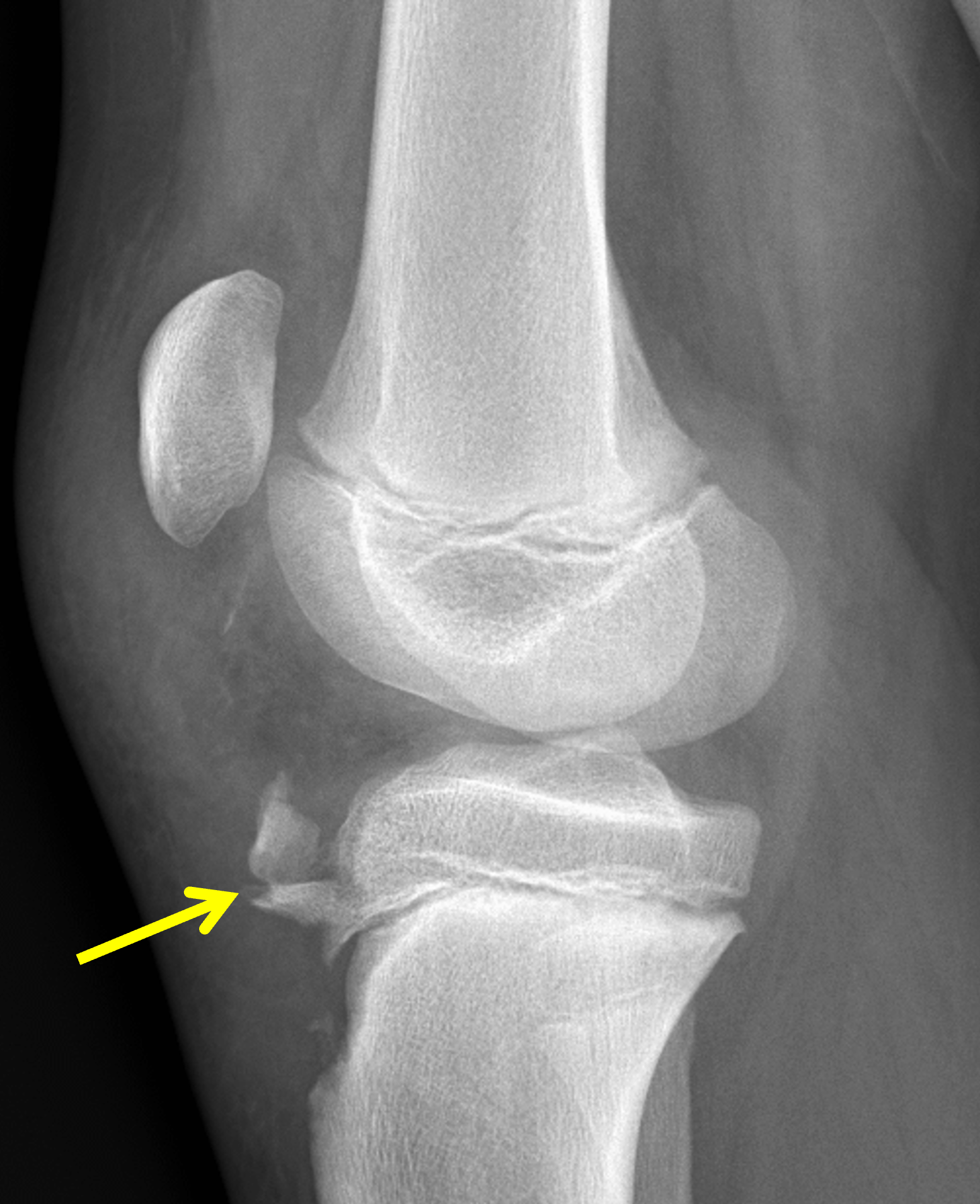
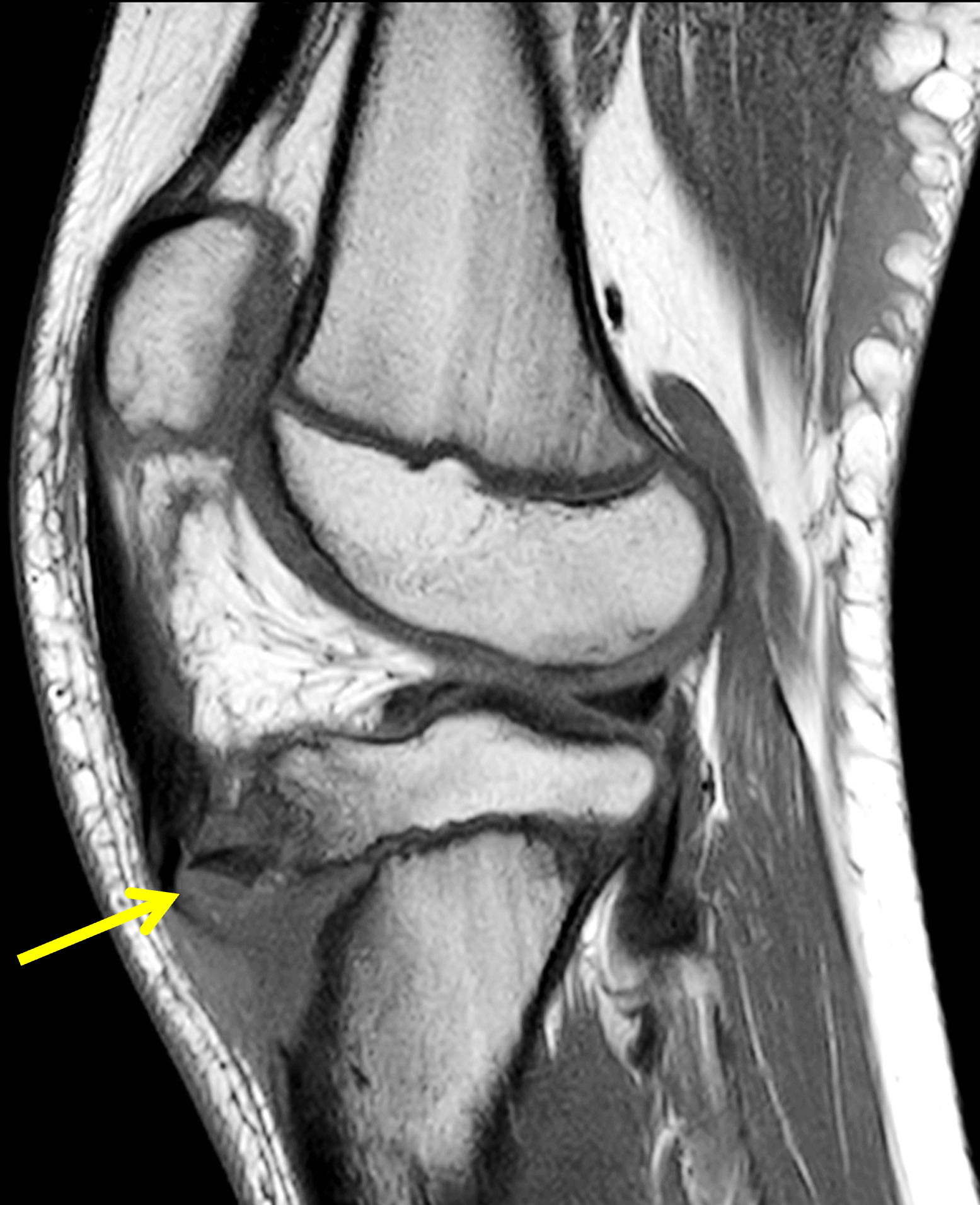


Figure 11. Acute tibial tuberosity avulsion fracture in a 12-year-old boy due to a football injury. (11A) Lateral radiograph shows a comminuted fracture of the anterior tibial tubercle apophysis (arrow) with the fracture fragments displaced cranially and rotated. Note the sharp borders of the fragments, unlike the ossicles in Osgood-Schlatter disease. Infrapatellar soft tissue swelling is present. (11B) Sagittal T1-weighted image shows one of the displaced and rotated fracture fragments (arrow). (11C) Sagittal fat-suppressed, fluid-sensitive image demonstrates marrow edema in the fracture fragment (arrow) and underlying bone (asterisk). (11D) Axial fat-suppressed, fluid-sensitive image confirms the fracture fragments (arrows) and marrow edema (asterisk). Note the disrupted anterior bone cortex.
Tendinosis represents a spectrum of pathology ranging from hypoxic and/or mucoid degeneration of the tendon to partial tears. Patellar tendinosis (also known as “jumper’s knee”) is due to overuse, typically in patients participating in sports that include running or jumping. The imaging findings are reviewed in a prior Web Clinic. Focal patellar tendinosis most commonly involves the proximal tendon; diffuse disease involving the entire tendon also occurs. Less commonly, the condition will be confined to the distal tendon (Figure 12), where symptoms can mimic those of Osgood-Schlatter disease.
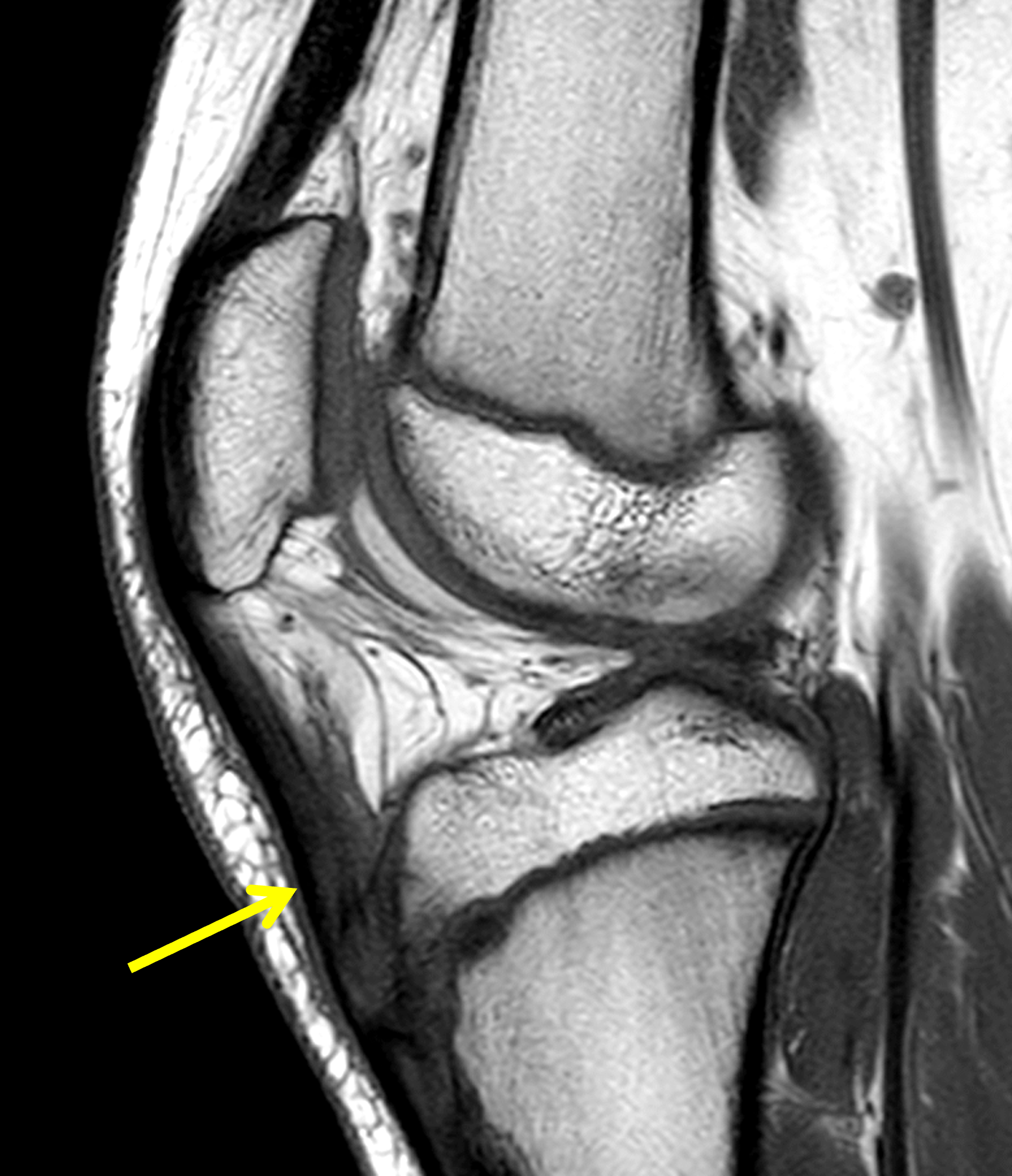

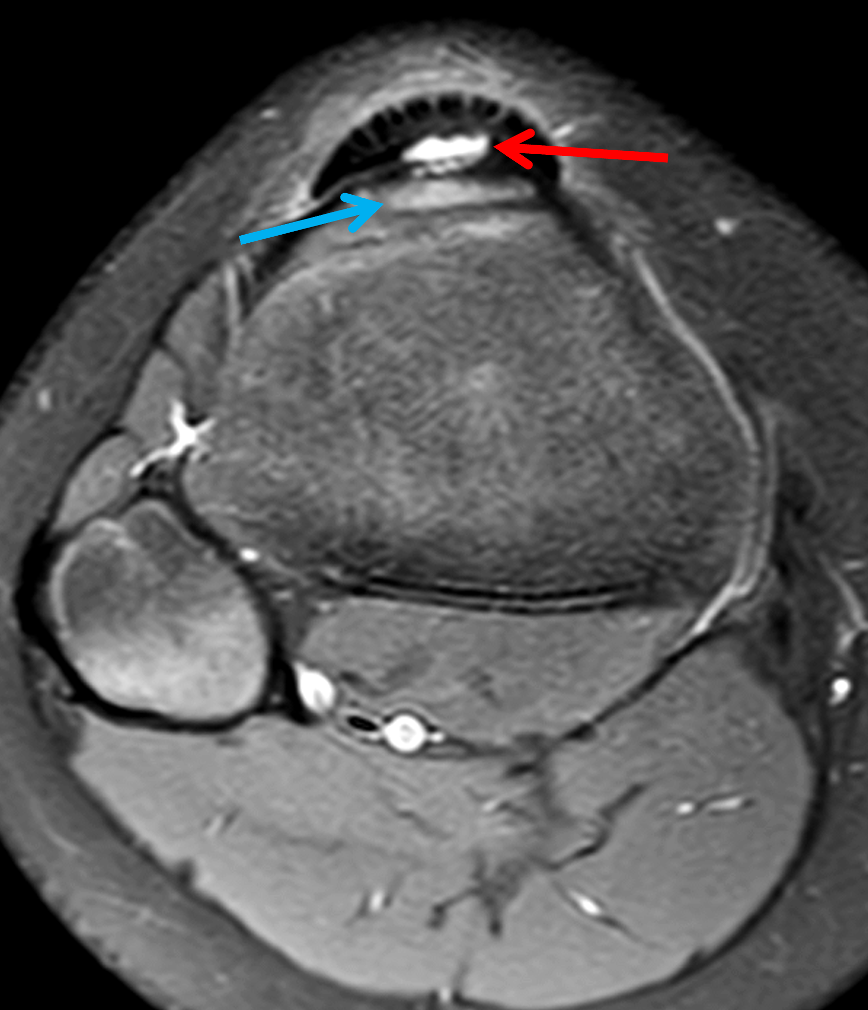
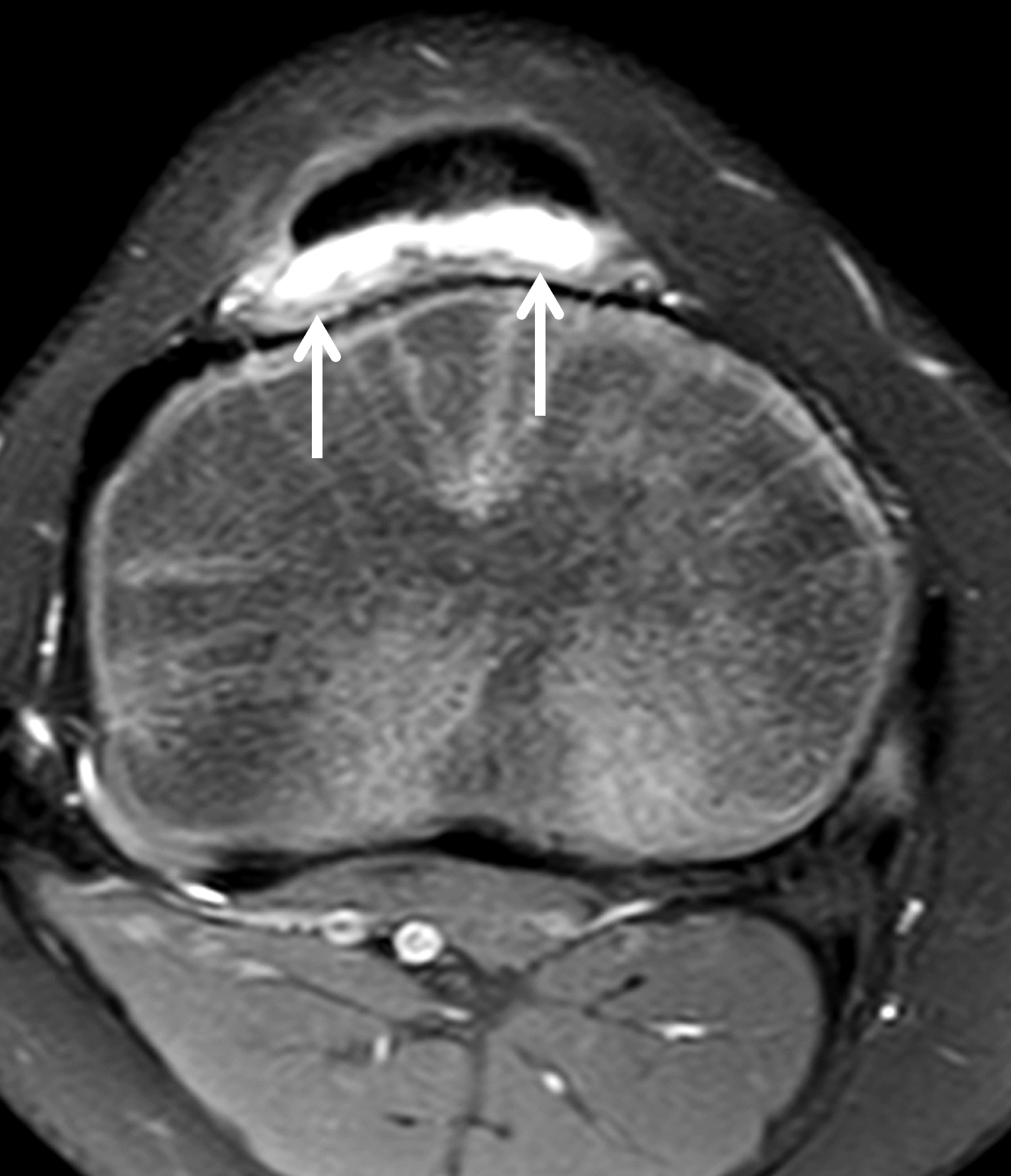
Figure 12: Distal patellar tendinosis and partial tendon tear in an 11-year-old boy who is active in sports. (12A) Sagittal T1-weighted image shows a thickened distal patellar tendon (arrow). (12B) Sagittal Fat-suppressed, fluid-sensitive image shows mucoid degeneration in the distal patellar tendon (yellow arrow) and a partial tear of the distal tendon insertion (red arrow). The deep infrapatellar bursa is mildly distended (white arrow). (12C) Axial fat-suppressed, fluid-sensitive image through the tibial tubercle shows a fluid cleft in the partly torn distal patellar tendon (red arrow). The tibial tuberosity apophysis (blue arrow) remains attached to the underlying bone, distinguishing the injury from an apophyseal avulsion. (12D) Axial fat-suppressed, fluid-sensitive image cranial to 12C demonstrates fluid in the deep infrapatellar bursa (arrows).
Traumatic, inflammatory, and overuse conditions of the infrapatellar (Hoffa’s) fat pad, as well as cysts or masses within the fat pad can also produce symptoms similar to Osgood-Schlatter disease. Disorders involving the fat pad are addressed in a prior Web Clinic as are infrapatellar ganglion cysts. Multiple bursa exist in the anterior knee, including superficial infrapatellar and deep infrapatellar bursae. Deep infrapatellar bursitis has been reported in pre-adolescent baseball players related to the amount of weekly practice (Figure 13).23 An adventitial bursa sometimes develops superficial to the anterior tibial tuberosity. Inflammation of any of these bursae – typically due to overuse, trauma, or inflammatory arthritis – can produce symptoms similar to Osgood-Schlatter disease.
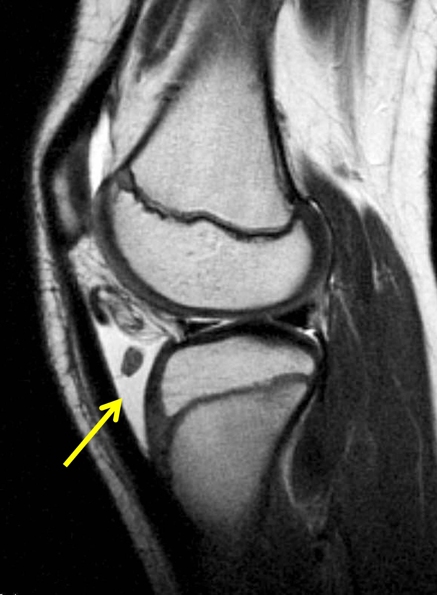
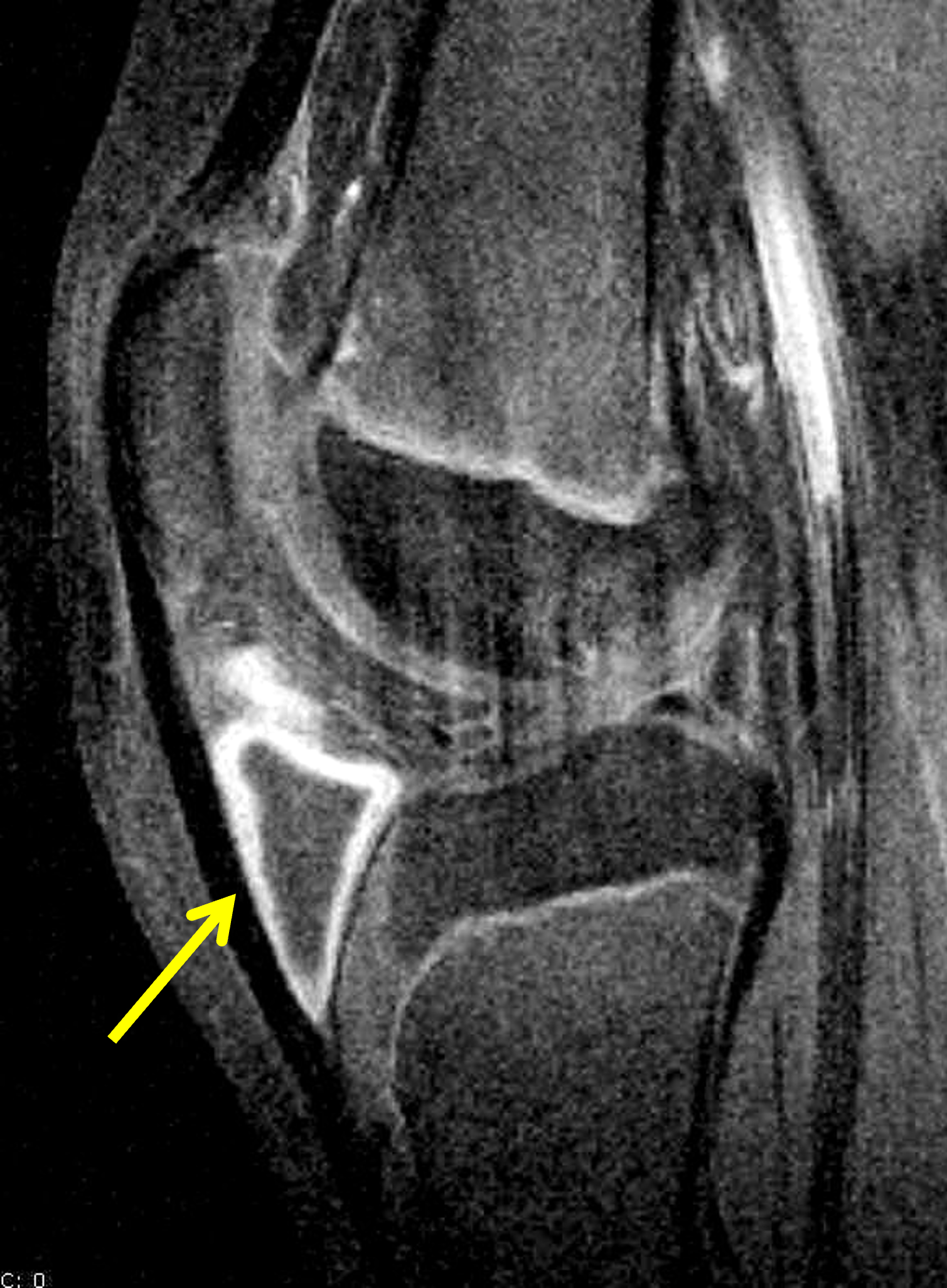
Figure 13. 10-year-old boy with anterior, infrapatellar pain due to deep infrapatellar bursitis. (13A) Sagittal T2-weighted image demonstrates fluid and debris within the deep infrapatellar bursa (arrow), in the caudal aspect of Hoffa’s fat pad. (13B) Sagittal fat-suppressed, T1-weighted image acquired following intravenous contrast administration shows enhancing synovium surrounding the distended bursa (arrow).
Other causes of anterior knee pain in an adolescent include acute patellar sleeve avulsion fractures and chronic Sinding-Larsen-Johansson syndrome, and disorders in the spectrum “patellofemoral pain syndrome,” including patellofemoral maltracking, instability, chondromalacia, and chondral trauma. However, the symptoms in these disorders are located more proximally (over the patellofemoral articulation) compared to the pain of Osgood-Schlatter disease and are usually well characterized and differentiated by a combination of clinical findings and MRI.24,25 These conditions can develop in association with Osgood-Schlatter disease, or may occur independently.
Uncommonly, tumors or infection of the proximal tibia may present similarly, though pain in these cases is often atypical and not activity related. Imaging in these cases is usually very helpful in making the correct diagnosis.26
Conclusion
Osgood-Schlatter disease, a traction apophysitis of the tibial tubercle, is a common cause of activity-related anterior knee pain and swelling in adolescents and preadolescents participating in sports that involve jumping, squatting, and lunging, often occurring during or following a recent growth spurt. The diagnosis can frequently be made clinically, but radiographs showing soft tissue swelling anterior to the tibial tubercle with or without bone changes in the anterior tibial tubercle are useful associated findings. However, a fragmented appearance of the apophysis may be normal before skeletal maturity and should not lead to a diagnosis of Osgood-Schlatter disease in the absence of symptoms. MRI may be useful to distinguish active disease – characterized by marrow edema in the anterior tibial tuberosity or adjacent ossicles, soft tissue edema in the inferior aspect of Hoffa’s fat pad and subcutaneous tissues, and/or infrapatellar bursitis – from inactive disease or sequela of previously healed Osgood-Schlatter disease. Treatment is usually conservative. Most patients fully recover, though with a variable course. Recent literature suggests an association with patellofemoral maltracking including in adults with a prior diagnosis of Osgood-Schlatter disease. Imaging studies obtained in patients with suspected Osgood-Schlatter disease should be evaluated for findings associated with patellofemoral maltracking, as well as other conditions that can cause anterior knee pain.
References
- Resnick D. Osteochondroses. In: Resnick D, editor. Diagnosis of Bone and Joint Disorders. 4th ed: W. B. Saunders; 2002. p. 3714–3718. ↩
- Lazerte GD, Rapp IH. Pathogenesis of Osgood-Schlatter’s disease. Am J Pathol. 1958;34(4):803–815. ↩
- Osgood RB. Lesions of the tibial tubercle occurring during adolescence. 1903. Clin Orthop Relat Res. 1993(286):4–9. ↩
- Rosenberg ZS, Kawelblum M, Cheung YY, Beltran J, Lehman WB, Grant AD. Osgood-Schlatter lesion: fracture or tendinitis? Scintigraphic, CT, and MR imaging features. Radiology. 1992;185(3):853–858. doi:10.1148/radiology.185.3.1438775 ↩
- Hirano A, Fukubayashi T, Ishii T, Ochiai N. Magnetic resonance imaging of Osgood-Schlatter disease: the course of the disease. Skeletal Radiol. 2002;31(6):334–342. doi:10.1007/s00256-002-0486-z ↩
- Ladenhauf HN, Seitlinger G, Green DW. Osgood-Schlatter disease: a 2020 update of a common knee condition in children. Curr Opin Pediatr. 2020;32(1):107–112. doi:10.1097/MOP.0000000000000842 ↩
- Nakase J, Goshima K, Numata H, Oshima T, Takata Y, Tsuchiya H. Precise risk factors for Osgood-Schlatter disease. Arch Orthop Trauma Surg. 2015;135(9):1277–1281. doi:10.1007/s00402-015-2270-2 ↩
- Neuhaus C, Appenzeller-Herzog C, Faude O. A systematic review on conservative treatment options for Osgood-Schlatter disease. Phys Ther Sport. 2021;49178–187. doi:10.1016/j.ptsp.2021.03.002 ↩
- Rathleff MS, Winiarski L, Krommes K, Graven-Nielsen T, Holmich P, Olesen JL, et al. Activity Modification and Knee Strengthening for Osgood-Schlatter Disease: A Prospective Cohort Study. Orthop J Sports Med. 2020;8(4):2325967120911106. doi:10.1177/2325967120911106 ↩
- Lucenti L, Sapienza M, Caldaci A, Cristo C, Testa G, Pavone V. The Etiology and Risk Factors of Osgood-Schlatter Disease: A Systematic Review. Children (Basel). 2022;9(6). doi:10.3390/children9060826 ↩
- Ndjonko LCM, Klein JH, Chakraborty Y, Kata S, Alinda A, Abuelenein I, et al. Treatments for Osgood Schlatter Disease: A Systematic Review of the Literature. Orthop J Sports Med. 2026;14(3):23259671251387354. doi:10.1177/23259671251387354 ↩
- Kamel SI, Kanesa-Thasan RM, Dave JK, Zoga AC, Morrison W, Belair J, et al. Prevalence of lateral patellofemoral maltracking and associated complications in patients with Osgood Schlatter disease. Skeletal Radiol. 2021;50(7):1399–1409. doi:10.1007/s00256-020-03684-6 ↩
- Guldhammer C, Rathleff MS, Jensen HP, Holden S. Long-term Prognosis and Impact of Osgood-Schlatter Disease 4 Years After Diagnosis: A Retrospective Study. Orthop J Sports Med. 2019;7(10):2325967119878136. doi:10.1177/2325967119878136 ↩
- Gholve PA, Scher DM, Khakharia S, Widmann RF, Green DW. Osgood Schlatter syndrome. Curr Opin Pediatr. 2007;19(1):44–50. doi:10.1097/MOP.0b013e328013dbea ↩
- Circi E, Atalay Y, Beyzadeoglu T. Treatment of Osgood-Schlatter disease: review of the literature. Musculoskelet Surg. 2017;101(3):195–200. doi:10.1007/s12306-017-0479-7 ↩
- Guszczyn T, Kulesza M, Maciag G, Kicman A, Lawicki S. The Effectiveness of Treating Osgood-Schlatter Disease (OSD) with Leukocyte-Rich Platelet-Rich Plasma (LR-PRP) Depending on the Duration of the Disease. J Clin Med. 2024;13(14). doi:10.3390/jcm13144220 ↩
- Guszczyn T, Kulesza M, Kicman A, Motyka J, Lawicki S. Enhanced Pain Relief and Function Improvement in Children with Osgood-Schlatter Disease: Leukocyte-Rich Platelet-Rich Plasma (LR-PRP) as a Complementary Treatment to Standard Conservative Therapy. Med Sci Monit. 2023;29e941523. doi:10.12659/MSM.941523 ↩
- Mun F, Hennrikus WL. Surgical Treatment Outcomes of Unresolved Osgood-Schlatter Disease in Adolescent Athletes. Case Rep Orthop. 2021;20216677333. doi:10.1155/2021/6677333 ↩
- Bosworth D. Autologous bone pegging for epiphysitis of the tibial tubercle. J Bone Joint Surg. 1934;16(4):829. ↩
- Weiss JM, Jordan SS, Andersen JS, Lee BM, Kocher M. Surgical treatment of unresolved Osgood-Schlatter disease: ossicle resection with tibial tubercleplasty. J Pediatr Orthop. 2007;27(7):844–847. doi:10.1097/BPO.0b013e318155849b ↩
- Jakob RP, von Gumppenberg S, Engelhardt P. Does Osgood–Schlatter disease influence the position of the patella? J Bone Joint Surg Br. 1981;63B(4):579–582. doi:10.1302/0301-620X.63B4.7298689 ↩
- Pan T, Mun F, Martinazzi B, King TS, Petfield JL, Hennrikus WL. The posterior tibial slope and Insall-Salvati index in operative and nonoperative adolescent athletes with Osgood-Schlatter disease. Arch Orthop Trauma Surg. 2022;142(12):3903–3907. doi:10.1007/s00402-021-04314-z ↩
- Omae H, Ohsawa T, Omodaka T, Hashimoto S, Oshima A, Takase R, et al. Deep Infrapatellar Bursitis in Preadolescent Baseball Players: A Cross-Sectional Study. Orthop J Sports Med. 2022;10(3):23259671221083584. doi:10.1177/23259671221083584 ↩
- Diederichs G, Issever AS, Scheffler S. MR imaging of patellar instability: injury patterns and assessment of risk factors. Radiographics. 2010;30(4):961–981. doi:10.1148/rg.304095755 ↩
- Watts RE, Gorbachova T, Fritz RC, Saad SS, Lutz AM, Kim J, et al. Patellar Tracking: An Old Problem with New Insights. Radiographics. 2023;43(6):e220177. doi:10.1148/rg.220177 ↩
- Jamshidi K, Mirkazemi M, Izanloo A, Mirzaei A. Benign bone tumours of tibial tuberosity clinically mimicking Osgood-Schlatter disease: a case series. Int Orthop. 2019;43(11):2563–2568. doi:10.1007/s00264-019-04397-9 ↩
