Radsource radiologists are constantly communicating and sharing knowledge with each other. In our blog series Today’s Interesting Case, our team will post notable cases and images for discussion from time to time.
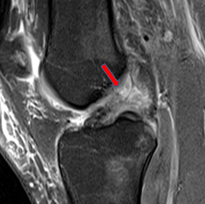
A fat suppressed proton density-weighted sagittal image reveals a proximal ACL disruption.
In today’s interesting case, an 18 year-old male presents following a recent skiing injury. The severity of the patient’s injury can be seen in the MR images finding three ligaments completely torn. Multi-ligamentous injuries such as in this case are strongly suggestive of knee dislocation.
As Dr. Eric Chang and Dr. Donald Resnick explain in the June 2011 web clinic on multi-ligamentous and tendon injuries suggesting knee dislocation, “In the medical literature, the term “knee dislocation” is commonly used to describe a knee either with multiple injured ligaments or with multi-directional instability, findings that can result from either knee subluxation or a complete knee dislocation1.”
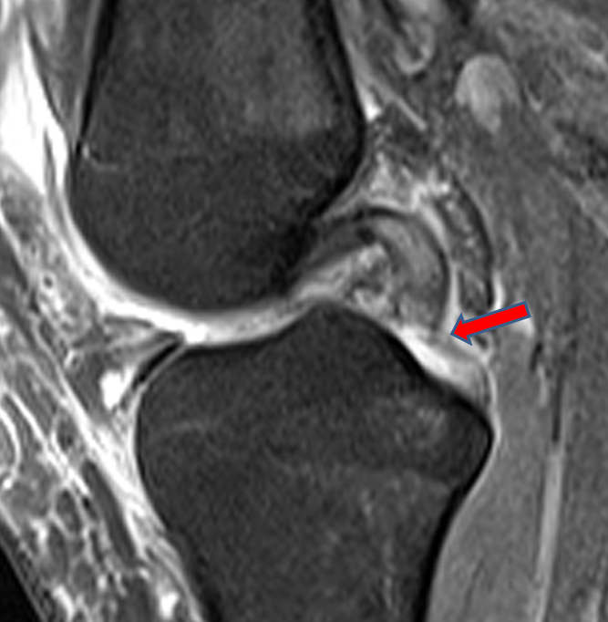
An adjacent sagittal slice demonstrate a complete distal PCL disruption as well.
Although knee dislocations are rare, their true prevalence is unknown because of missed diagnoses or misdiagnosis. The prevalence has been reported to be as high as 0.11% in one series that reviewed over 17,000 consecutive knee MRI examinations2. Most commonly knee dislocations are associated with high-velocity trauma related to motor vehicle accidents1; however, in some reports low-velocity injuries from sports activities have been the predominate cause of knee dislocation2,3.”
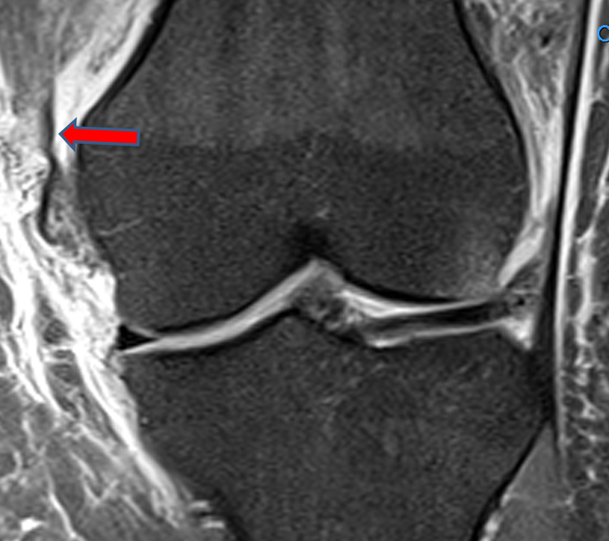
A fat-suppressed proton density-weighted coronal image demonstrates a Grade III tear of the medial collateral ligament complex. The tibial collateral is disrupted and flipped superiorly.
Treatment for knee dislocations has advanced greatly over the years thanks to modern surgical techniques4 and prompt MRI assisted diagnosis. “Prior to surgery MRI allows accurate characterization of the extent of injuries to supporting structures of the knee, as well as identification of meniscal tears which may require partial meniscectomy or repair. MR thus provides a valuable roadmap towards determining the appropriate surgical approach,” state Dr. Chang and Dr. Resnick.
References
1 Brautigan, B. and D.L. Johnson, The epidemiology of knee dislocations. Clin Sports Med, 2000. 19(3): p. 387-97.
2 Bui, K.L., et al., Knee dislocations: a magnetic resonance imaging study correlated with clinical and operative findings. Skeletal Radiol, 2008. 37(7): p. 653-61.
3 Shelbourne, K.D., M.S. Haro, and T. Gray, Knee dislocation with lateral side injury: results of an en masse surgical repair technique of the lateral side. Am J Sports Med, 2007. 35(7): p. 1105-16.
4. Taylor, A.R., G.P. Arden, and H.A. Rainey, Traumatic dislocation of the knee. A report of forty-three cases with special reference to conservative treatment. J Bone Joint Surg Br, 1972. 54(1): p. 96-102. – See more at: https://radsource.us/multi-ligamentous-and-tendon-injury/#sthash.QFxfHMfB.dpuf
