Radsource radiologists are constantly communicating and sharing knowledge with each other. In our blog series Today’s Interesting Case, our team will post notable cases and images for discussion from time to time.
A 36 year old male suffered a twisting injury 2.5 weeks ago.
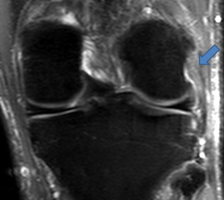
A coronal fat-suppressed proton density-weighted image reveals a thickened and edematous proximal fibular collateral ligament.
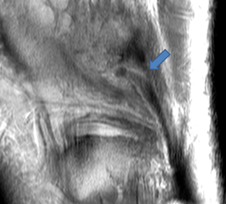
A sagittal proton density-weighted image demonstrates focal transverse disruption of the proximal fibular collateral ligament.
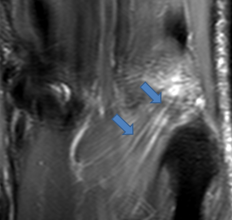
A posterior fat-suppressed proton density-weighted coronal image reveals edema and fluid along the course of the popliteus muscle. Edema was also present at the popliteus origin (not shown).
The ACL and PCL were normal in this patient. The findings are compatible with an isolated posterolateral corner injury, an uncommon occurrence. For more on this topic, see Dr. Michael Stadnick’s web clinic on posterolateral corner injury.
