Radsource radiologists are constantly communicating and sharing knowledge with each other. In our blog series Today’s Interesting Case, our team will post notable cases and images for discussion from time to time.
Our case today involves rare pathology. The patient is a 51 year-old female presenting with medial ankle pain on weight-bearing. She experienced trauma with a fracture of the medial malleolus 5 months earlier.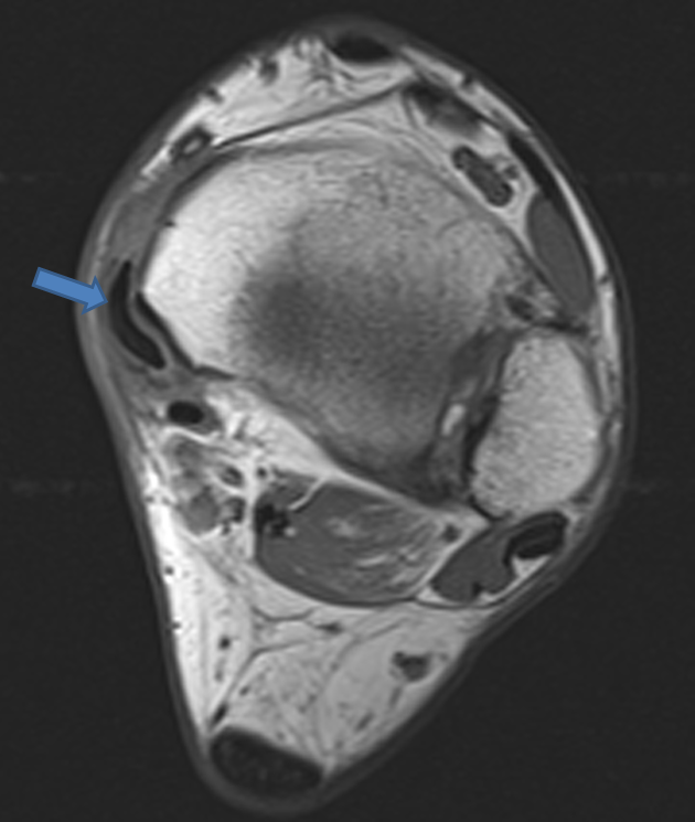
A T1-weighted axial image reveals anteromedial subluxation of the posterior tibial tendon (arrow).
As explained by Dr. Pamela Burdett in her web clinic on posterior tibial tendinopathy, subluxation or dislocation of the posterior tibial tendon is a rare event, “Dislocation of the posterior tibial tendon is rare, and thought to be traumatic. It is usually associated with a torn flexor retinaculum, which allows the tendon to slip out of the retromalleolar groove. An avulsion fracture at the medial malleolar attachment of the flexor retinaculum may be seen in such cases1.
“MRI is useful in evaluating the full range of posterior tibial tendon dysfunction, including tenosynovitis, tendon tears, and pes planus deformity. The ability of MR to judge the extent of disease and associated abnormalities such as spring ligament tears is important in surgical planning,” states Burdett.
- Schweitzer ME, Karasick D MR Imaging of Disorders of the Posterior Tibialis Tendon Am. J. Roentgenol., Sep 2000; 175: 627 – 635. ↩
