Radsource radiologists are constantly communicating and sharing knowledge with each other. In our blog series Today’s Interesting Case, our team will post notable cases and images for discussion from time to time.
A 24 year-old male presents with knee pain and swelling and a posterior mass.
A fat-suppressed proton density-weighted sagittal image reveals extensive lobulated soft tissue thickening within the joint, obscuring the ACL. Lobulated soft tissue with similar signal characteristics is see posterior to the joint.
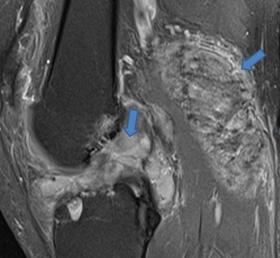
A fat-suppressed proton density-weighted sagittal image reveals extensive lobulated soft tissue thickening within the joint, obscuring the ACL. Lobulated soft tissue with similar signal characteristics is see posterior to the joint.
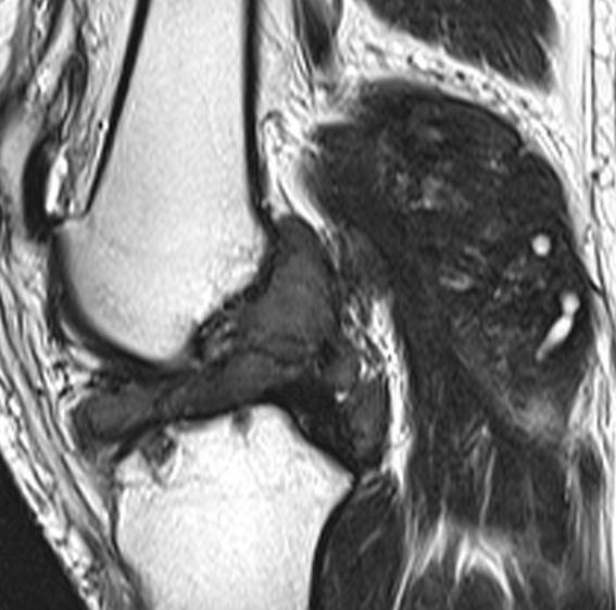
On a T2-weighted sagittal view, regions of very low signal intensity are more readily apparent within the lesions, likely due to the presence of hemosiderin.
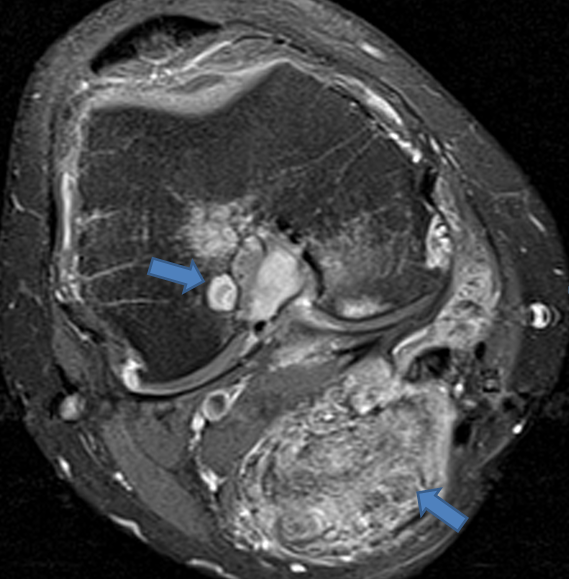
On a fat-suppressed proton density-weighted axial view, an osseous erosion is apparent, and the posterior “mass” is seen to be secondary to the process filling a large popliteal cyst.
The findings are compatible with a severe synovial proliferative process, in this case due to an unusually severe case of pigmented villonodular synovitis (PVNS).
The only curative treatment for PVNS is surgery, and in such cases, MRI provides valuable preoperative information regarding the extent of the process.
Find more information on this topic, see Dr. Mark Awh’s Pigmented Villondular Synovitis Web Clinic from March 2005.
