Radsource radiologists are constantly communicating and sharing knowledge with each other. In our blog series Today’s Interesting Case, our team will post notable cases and images for discussion from time to time.
For our case today, we present an 18 year-old male with knee pain and stiffness after a fall down steps.
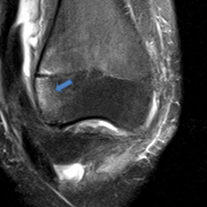
A fat-suppressed proton density-weighted coronal image reveals a bone bruise at the anterolateral aspect of the lateral femoral condyle, typical for recent patellar dislocation.
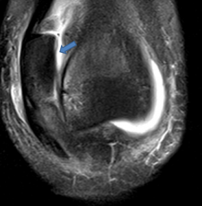
On a more anterior coronal image, the dislocated patella is identified.
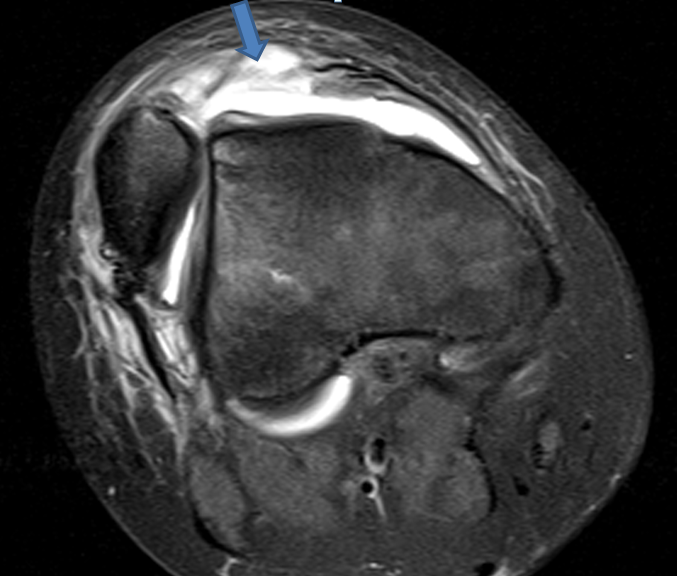
The persistent patellar dislocation is confirmed on an axial image. Complete disruption of the medial retinaculum is also apparent.
This case is unusual because it is exceedingly rare to image a patient with the patella persistently dislocated, as almost all patellar dislocations spontaneously reduce when the patient extends the knee. For more on patellar dislocation, see Dr. Lisa Ballehr’s Transient Lateral Patellar Dislocation web clinic from April 2013.