Radsource radiologists are constantly communicating and sharing knowledge with each other. In our blog series Today’s Interesting Case, our team will post notable cases and images for discussion from time to time.
Our case today explores the use of magnetic resonance imaging (MR) in the evaluation of the affected hip in patients who have undergone hip arthroplasty.
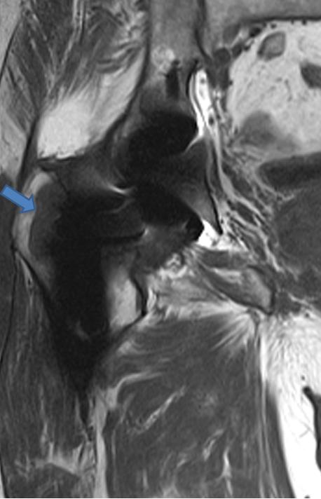
A 74 year-old male with a history of prior hip arthroplasty presents with a several week history of right hip pain and swelling. A T1-weighted coronal image reveals osteolysis at the greater trochanter.
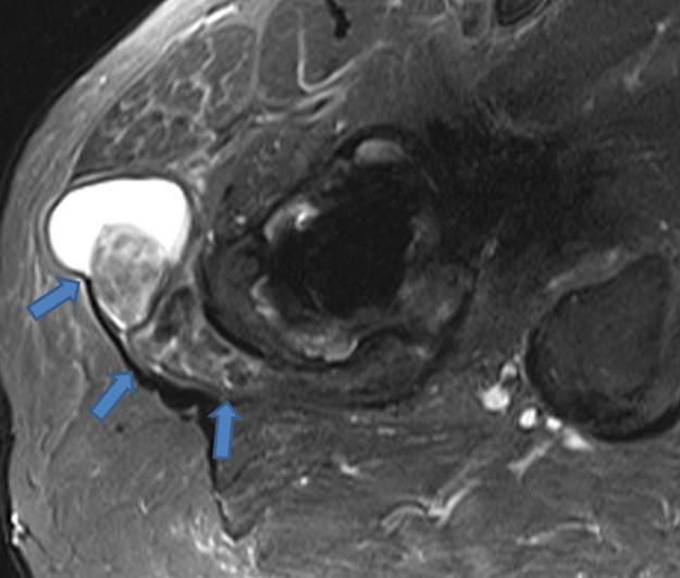
A STIR axial image demonstrates a complex fluid collection extending through the posterolateral joint capsule compatible with a pseudotumor. Areas of low signal intensity within the fluid are presumably related to metallic debris. The findings in this case are typical for particle disease.
Dr. Mark Awh’s 2011 web clinic on hip arthroplasty goes into more depth on the MR imaging technique and diagnosis of particle disease (including including synovitis, osteolysis, and pseudotumor formation).
