
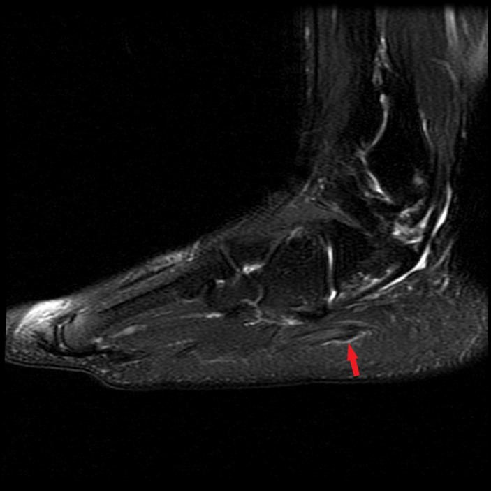
Fat-suppressed2-weighted (20A) and T1-weighted (20B) images in the coronal plane and a sagittal fat-suppressed T2-weighted image (20C) of the ankle in a 47-year-old female with lateral heel pain demonstrates focal nodular intermediate signal thickening along the lateral cord of the plantar aponeurosis proximal to the insertion suggestive of a plantar fibroma (arrows). This patient also had chronic denervation of the abductor digiti minimi muscle (asterisk).


