
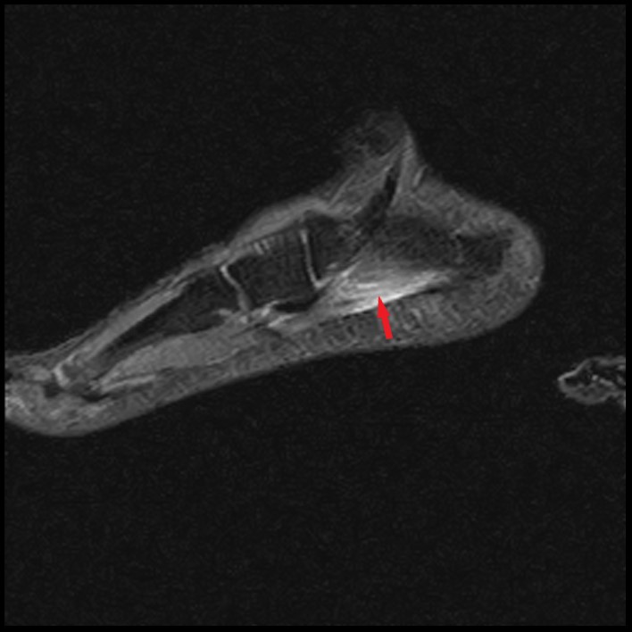
Coronal (19A) and sagittal (19B) fat-suppressed T2-weighted images in a different patient with acute lateral heel and midfoot pain demonstrate edema within the abductor digiti muscle that extends along the myotendinous junction (arrows) suggestive of myotendinous strain rather than diffusely involving the muscle as in subacute denervation. Coronal T1-weighted image (19Cc) demonstrates preserved muscle bulk of the abductor digiti minimi muscle (asterisk) without fatty infiltration.


