
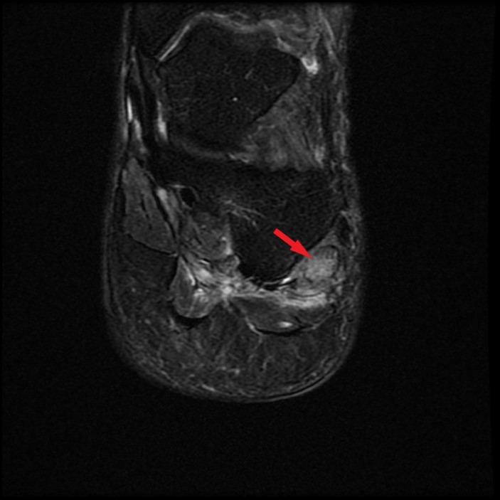
Axial (12A), coronal (12B), and sagittal (12C) fat-suppressed T2-weighted images in a 53-year-old with lateral midfoot pain after twisting injury 5 weeks ago demonstrates a hypertrophied, edematous os peroneum (arrows) located near the level of the calcaneocuboid joint within the peroneus longus tendon. The adjacent peroneus longus tendon (arrowheads) is moderately tendinopathic between the os peroneum and the cuboid tunnel. Oblique radiograph of the foot (12D) shows the corresponding enlarged os peroneum (arrow) with mild irregularity along the distal margin.


