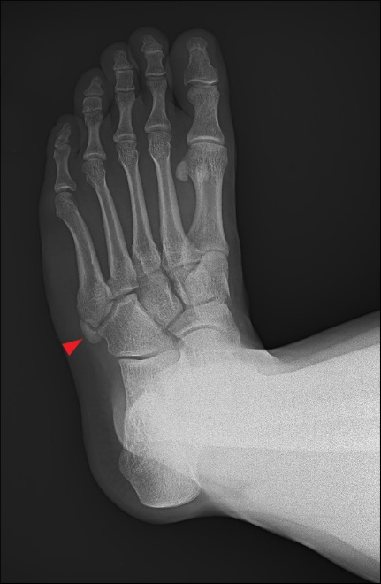
Sagittal proton density-weighted image (11A) and axial fat-suppressed proton density-weighted image (11B) in a 21-year-old male with chronic pain at the base of the fifth metatarsal demonstrates a hypertrophied, triangular-shaped ossicle (asterisks) adjacent to the fifth metatarsal base. Edema surrounds the synchondrosis (arrows) suggestive of micromotion. Lateral cord of the plantar aponeurosis (arrowhead) inserts on the ossicle. Oblique radiograph of the foot (11C) demonstrates the accessory ossicle at the fifth metatarsal base (arrowhead).


