
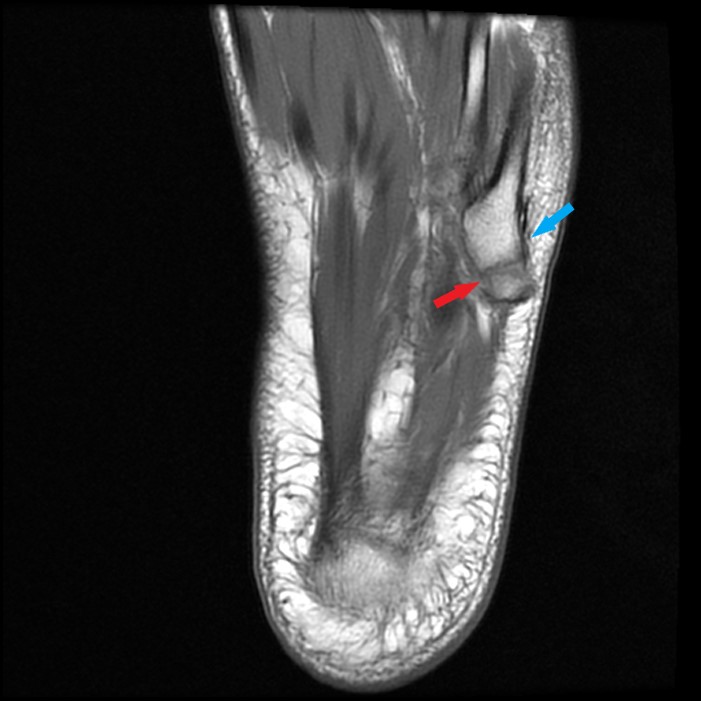
Sagittal (10A) and axial (10B) T1-weighted images in another 61-year-old female with lateral midfoot pain demonstrates a mildly displaced avulsion fracture of the fifth metatarsal base tuberosity (arrows). Oblique coronal fat-suppressed proton density-weighted image (10C) demonstrates bone marrow edema associated with the fracture and the adjacent insertion of the lateral cord of the plantar fascia (arrowhead). Note the peroneus brevis insertion spans the fracture (blue arrow) and thus is unlikely to be the primary contributing factor to the avulsion fracture.


