
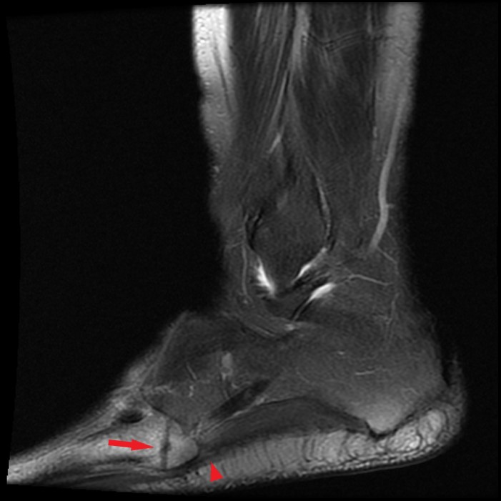
Sagittal T1-weighted (9A), fat-suppressed T2-weighted (9B) images and axial T1-weighted (9C) images of the ankle in a 48-year-old male with 1 month history of pain localized to the base of the fifth metatarsal. A transverse fracture (arrows) is noted through the fifth metatarsal tuberosity with surrounding bone marrow edema. Note the location of the insertion of the lateral cord of the plantar fascia on the plantar lateral aspect of the tuberosity (arrowheads).


