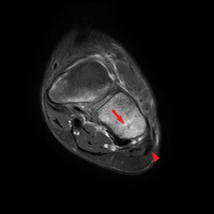
Coronal fat-suppressed T2-weighted image (8A), sagittal (8B) and axial (8C) T1-weighted images in 11-year-old male with 3-week history of lateral midfoot pain. Fracture line noted in the lateral aspect of the cuboid on the T2 and T1-weighted images (arrows) with surrounding marrow edema. Note the proximity of the cuboid fracture to the lateral cord of the plantar fascia located just plantar and lateral to the cuboid (arrowheads).
Categories
Our Mission
To provide the highest quality clinical and technology services to customers and patients, in the spirit of continuous improvement and innovation.
Sign up for our monthly web clinics![badge](https://assets.capterra.com/badge/9aad5659fd7efbbfa1a7ad0918d623ad.png?v=2117372&p=168760)
Categories
Helpful Links
Get in Touch
Radsource, LLC
750 Old Hickory Blvd, Suite 1-260
Brentwood, TN 37027
ph 615.376.7502
[email protected]