
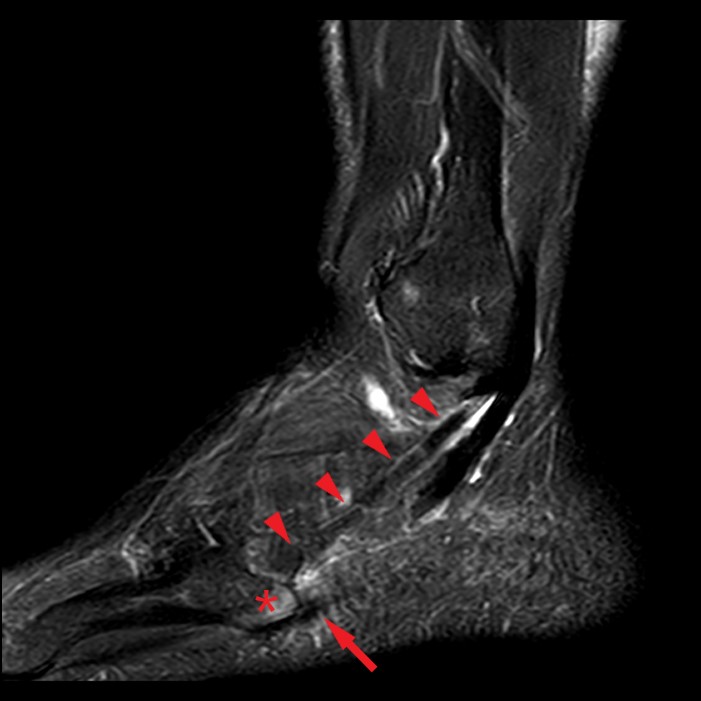
Fat-suppressed T2-weighted axial (6A), coronal fat-suppressed proton density-weighted (6B), and sagittal fat-suppressed T2-weighted (6C) images in another 51-year-old male with acute on chronic midfoot pain near the cuboid tunnel. Moderate thickening and edema along the distal lateral cord of the plantar aponeurosis (arrows) with mild insertional bone marrow edema at the fifth metacarpal base attachment (asterisks) is present on the fat-suppressed T2-weighted images.


