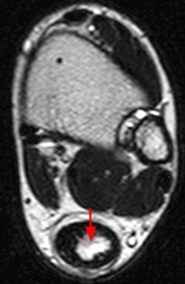
10B – Fat-suppressed T2-weighted sagittal (10a) and T2-weighted axial (10b) images from a 47-year-old male who presented with a history of a palpable Achilles nodule that arose after a fall one year earlier. The sagittal image reveals fusiform swelling of the tendon compatible with chronic tendinosis. A fluid-filled interstitial tear is present within the mid tendon (arrows) on both images. The history and clinical presentation were not consistent with infection.


