
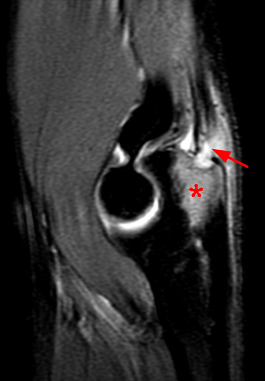
3A – Fat-suppressed proton density-weighted sagittal (3a) and fat-suppressed T2-weighted coronal (3b) images of the elbow in a 29-year-old male who sustained a fall with an open wound. Focal marrow edema (asterisks) is apparent within the olecranon, later confirmed to be due to osteomyelitis. Adjacent fluid signal intensity is present within the distal triceps tendon (arrows), compatible with a small intratendinous abscess.


