
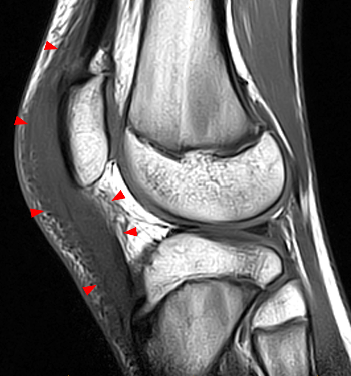
2A – Anterior swelling and edema are apparent on the T1-weighted (2a) and fat-suppressed proton density-weighted (2b) sagittal images (arrowheads). Central fluid signal intensity is present within the patellar tendon (arrows) on the fat-suppressed proton density-weighted image.


