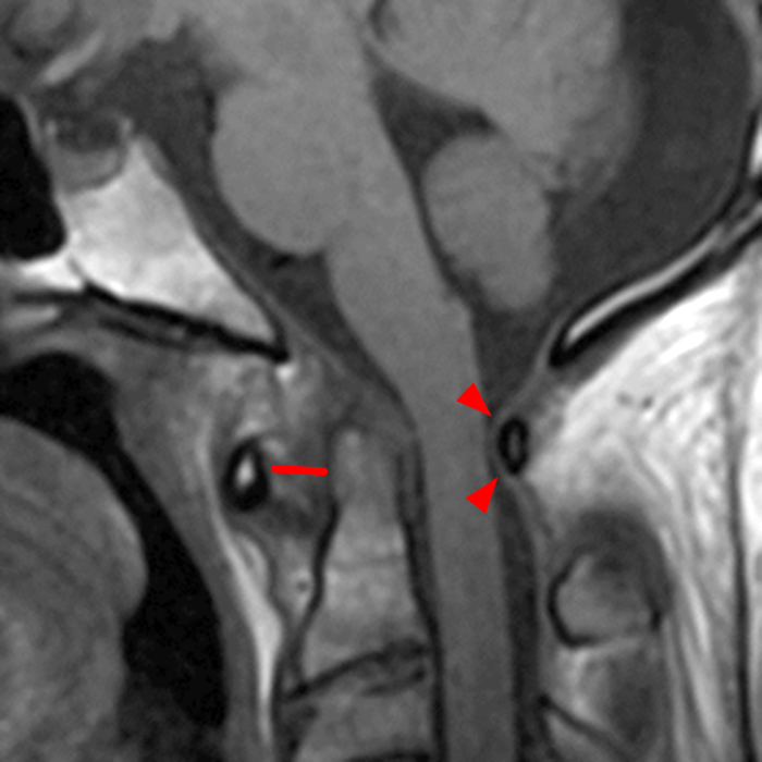
7A: Sagittal T1-weighted image demonstrates an increased atlantodental interval to 7 mm (lines) associated with laxity of the transverse ligament (short arrows).

7A: Sagittal T1-weighted image demonstrates an increased atlantodental interval to 7 mm (lines) associated with laxity of the transverse ligament (short arrows).
To provide the highest quality clinical and technology services to customers and patients, in the spirit of continuous improvement and innovation.
Sign up for our monthly web clinics750 Old Hickory Blvd, Suite 1-260
Brentwood, TN 37027
ph 615.376.7502
[email protected]


