
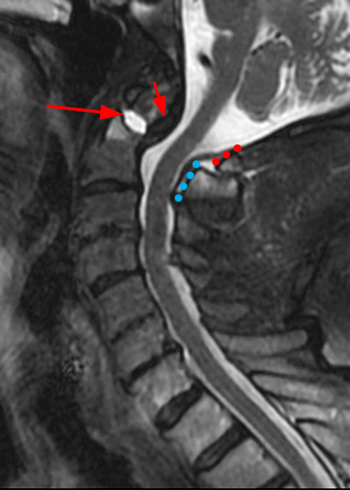
6B: STIR image shows a subacute Type II odontoid fracture with posterior displacement and diastasis at the fracture site (arrows), thickening of the transverse ligament (short arrows) and posterior translation of the C1 posterior arch (red dotted line) relative to the posterior arch of C2 (blue dotted line).


