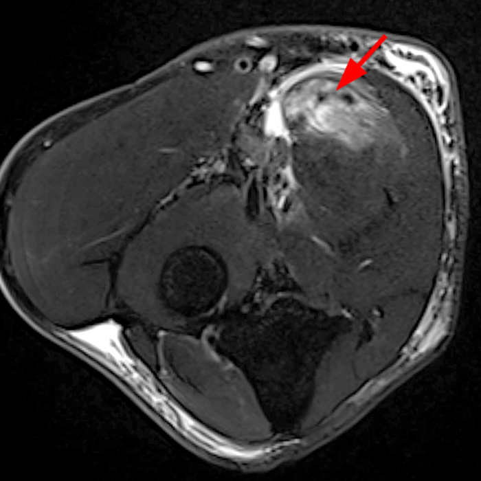
Figure 11: Axial T2-weighted fat-suppressed image demonstrates a focal myotendinous partial tear of the pronator teres (arrow) with focal muscle edema and partial myotendinous disruption.

Figure 11: Axial T2-weighted fat-suppressed image demonstrates a focal myotendinous partial tear of the pronator teres (arrow) with focal muscle edema and partial myotendinous disruption.
To provide the highest quality clinical and technology services to customers and patients, in the spirit of continuous improvement and innovation.
Sign up for our monthly web clinics750 Old Hickory Blvd, Suite 1-260
Brentwood, TN 37027
ph 615.376.7502
[email protected]


