
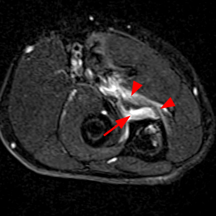
7A: Axial T2-weighted fat-suppressed (7A) and sagittal STIR (7B) images demonstrate a partial tear of the distal brachialis insertion with a small fluid gap at the tendinous insertion (arrow), mild subjacent marrow edema (asterisk) and edema in the remainder of the musculotendinous/musculoaponeurotic insertion (arrowheads).


