
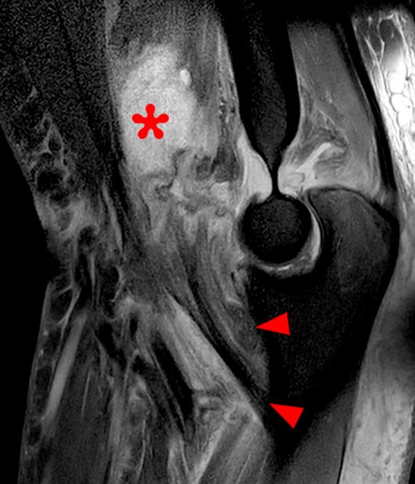
2B: Fat-suppressed axial T2-weighted (2A) and sagittal proton density-weighted (2B) images demonstrate a large brachialis intramuscular defect (asterisks) with diffuse surrounding muscle edema and heterogeneous fluid signal in the muscle gap. Note that the distal insertion of the brachialis onto the ulna is intact (arrowheads).


