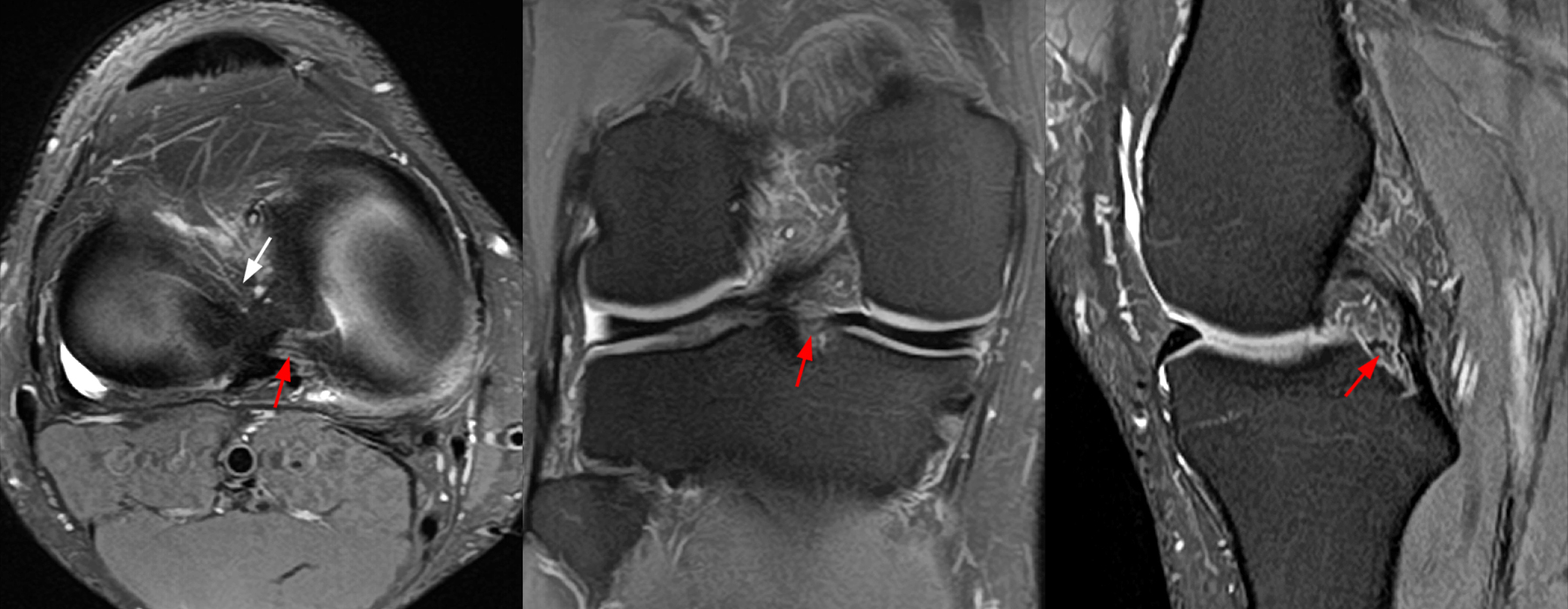
4A – Axial, coronal, and sagittal fat-suppressed proton density-weighted images demonstrate the normal striated appearance of the posterior root medial meniscus (red arrows). The anterior root lateral meniscus is also evident (white arrow).

4A – Axial, coronal, and sagittal fat-suppressed proton density-weighted images demonstrate the normal striated appearance of the posterior root medial meniscus (red arrows). The anterior root lateral meniscus is also evident (white arrow).
To provide the highest quality clinical and technology services to customers and patients, in the spirit of continuous improvement and innovation.
Sign up for our monthly web clinics750 Old Hickory Blvd, Suite 1-260
Brentwood, TN 37027
ph 615.376.7502
[email protected]


