
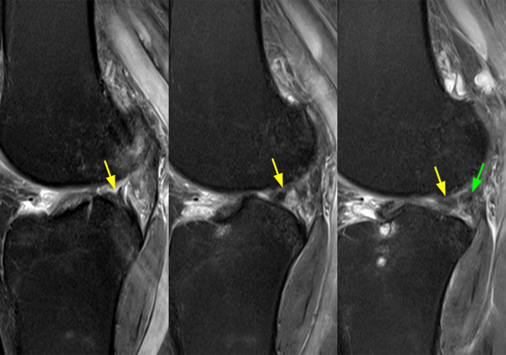
2D – Three contiguous sagittal fat-suppressed proton density-weighted images from medial to lateral demonstrate posterior horn lateral meniscus edema, fraying, and irregular contour adjacent to the root insertion (yellow arrows) with an intact meniscofemoral ligament (green arrow).


