
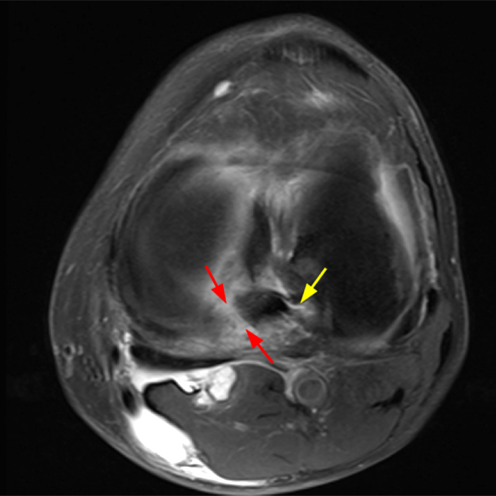
2A – An axial fat-suppressed proton density-weighted image demonstrates a full-thickness hyperintense cleft along the posterior horn medial meniscus adjacent to the root insertion (red arrows) and abnormally increased T2 signal in the posterior horn lateral meniscus adjacent to the root insertion (yellow arrow).


