Radsource radiologists are constantly communicating and sharing knowledge with each other. In our blog series Today’s Interesting Case, our team will post notable cases and images for discussion from time to time.
A 58 year-old male presents after acute injury from playing kickball 2 weeks ago. The patient felt a pop at the posterior thigh.
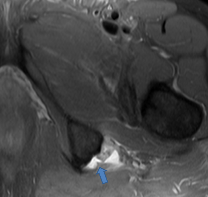
A fat-suppressed proton-density weighted image reveals a fluid filled gap at the expected hamstring origin from the ischial tuberosity.
.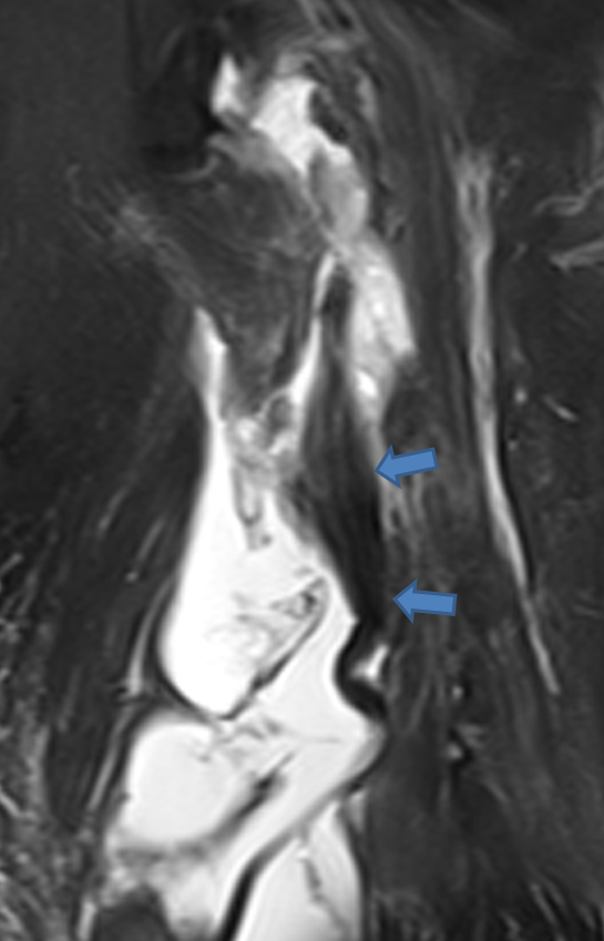
A fat-suppressed T2-weighted coronal image reveals the retracted hamstring tendons with surrounding fluid. The tendons are retracted approximately 5 cm from the origin
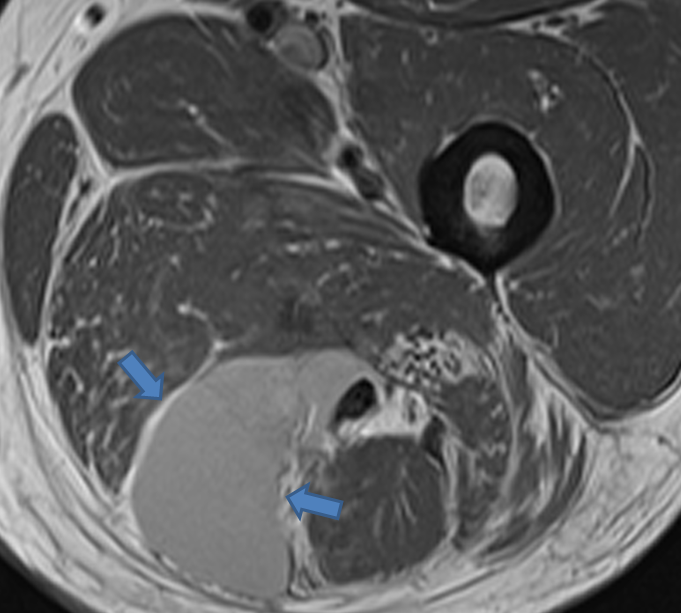
A corresponding T1-weighted axial view reveals that the fluid is hyperintense on T1, compatible with subacute hemorrhage.
This case demonstrates a particularly good example of a Grade III hamstring tear with an associated hematoma. As Dr. Michael Stadnick writes in the Hamstring Tears web clinic, “Grade III injuries represent a complete disruption of the musculotendinous junction. Retraction and laxity of the musculotendinous elements are seen. MRI is helpful in determining the extent of retraction in these injuries, an important feature for surgical planning.” Given the length of tendon retraction, surgery is likely indicated in this patient.
