Radsource radiologists are constantly communicating and sharing knowledge with each other. In our blog series Today’s Interesting Case, our team will post notable cases and images for discussion from time to time.
Today’s case involves a 46 year-old male who was injured playing hockey and presents with pain, bruising, and a lump in the anterior chest.
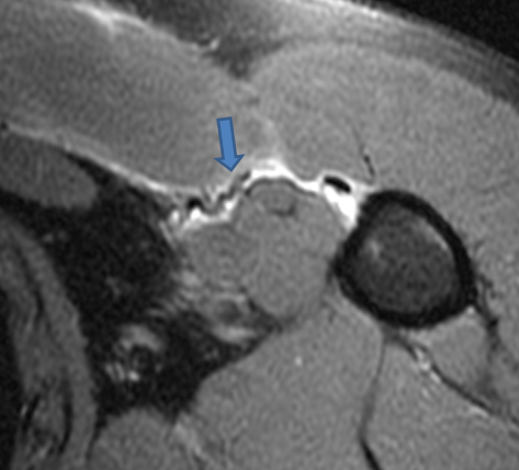
A fat-suppressed proton density weighted axial image reveals complete disruption of the sternal head tendon of the pectoralis major from the humeral attachment. The retracted tendon end is readily apparent (arrow).
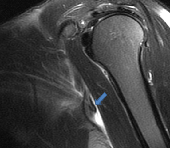
A fat-suppressed T2-weighted oblique coronal view confirms the torn and retracted sternal head tendon, with its distal end flipped cephalad.
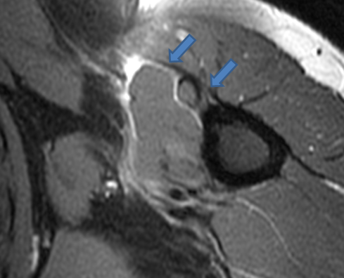
A more distal axial image reveals that the tendon attachment of the clavicular head of the pectoralis major remains intact.
This case nicely illustrates a common pattern of injury of the pectoralis. In our experience, if only one component of the pectoralis is injured, it is most commonly the sternal head. This pattern of injury is also important as recognizing a distal avulsion is important in operative planning. Avulsions from the humeral attachment are typically easier to repair than those that involve the musculotendinous junction.
For more information on this topic, see Dr. Michael Stadnick’s Pectoralis Tear Web Clinic from September 2006.