Radsource radiologists are constantly communicating and sharing knowledge with each other. In our blog series Today’s Interesting Case, our team will post notable cases and images for discussion from time to time.
A 15 year-old male presents following a direct blow to the shoulder in football. The surgeon suspects a labral tear.
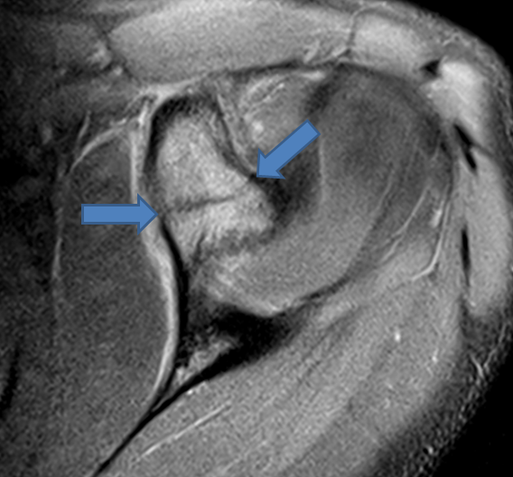
A fat-suppressed T2-weighted coronal oblique image reveals intense marrow edema within the coracoid process. The AC joint is unremarkable and other images revealed no labral tear.
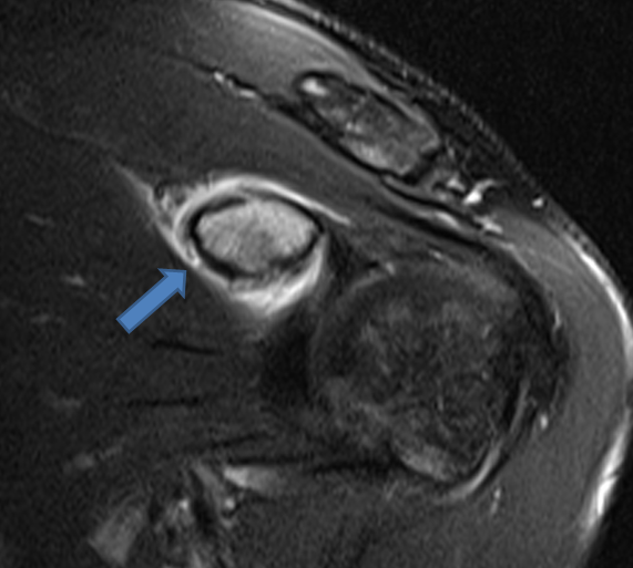
An acute transverse fracture of the coracoid process is revealed on the fat-suppressed proton density-weighted axial view.
Today’s case depicts a relatively rare injury, an isolated coracoid fracture. These typically occur due to direct trauma. Coracoid fractures are commonly associated with other injuries such as AC joint separation or clavicle fractures, which were not present in this patient. Non-displaced isolated coracoid fractures such as in this case are challenging to diagnose clinically or via plain radiographs. The diagnosis on MRI, however, is straightforward.